Search results (80 results)
-
 Aggressive Posterior Retinopathy of Prematurity
Oct 9 2012 by Audina M. Berrocal, MD FASRS
Aggressive posterior Type 1 ROP with bleeding from regression of the posterior hyaloid artery
Photographer: Ditte Hess CRA, BPEI
Imaging device: RETCAM
Condition/keywords: retinopathy of prematurity (ROP)
-
 Aggressive Posterior Retinopathy of Prematurity with Macular Hemorrhage
Oct 9 2012 by Audina M. Berrocal, MD FASRS
Aggressive posterior Type 1 ROP
Photographer: Ditte Hess CRA, BPEI
Imaging device: RETCAM
Condition/keywords: aggressive posterior retinopathy of prematurity (APROP), macular hemorrhage, retinopathy of prematurity (ROP)
-
 Aggressive Posterior Retinopathy of Prematurity with Macular Hemorrhage
Oct 9 2012 by Audina M. Berrocal, MD FASRS
APROP with multiple pre-retinal hemorrhages
Photographer: Ditte Hess CRA, BPEI
Imaging device: RETCAM
Condition/keywords: macular hemorrhage, retinopathy of prematurity (ROP)
-
 Aggressive Posterior Retinopathy of Prematurity
May 8 2017 by Juan Romo-Aguas
Fundus photograph of an 1 month and 21 days female with bilateral aggresive posterior retinopathy of prematurity.
Photographer: Juan C. Romo-Aguas, Asociación Para Evitar la Ceguera en México
Imaging device: Optos Daytona Ultra-widefield Retinal Imaging
Condition/keywords: aggressive posterior retinopathy of prematurity (APROP), retinopathy of prematurity (ROP)
-
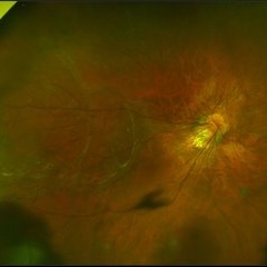
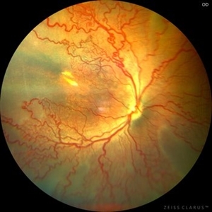 Aggressive Posterior Retinopathy of Prematurity (APROP)
May 16 2025 by KANWALJEET HARJOT MADAN, M.S. (Ophthalmology); FAICO (Vitreous - Retina)
This is the fundus picture of right eye of a premature neonate depicting Aggressive Posterior Retinopathy of Prematurity (APROP). It is a severe rapidly progressing form of retinopathy that can lead to vision loss and blindness. It requires prompt diagnosis and treatment in the form of anti-VEGF agents and laser photocoagulation.
Photographer: Dr. Kanwaljeet Harjot Madan, Thind Eye Hospital, Jalandhar City (Punjab) INDIA.
Imaging device: Zeiss Clarus
Condition/keywords: Oxygen Exposure, retinopathy of prematurity (ROP)
-
 AP-ROP
May 6 2017 by Juan Romo-Aguas
Aggressive posterior retinopathy of prematurity.
Condition/keywords: aggressive posterior retinopathy of prematurity (APROP), retinopathy of prematurity (ROP)
-
 APROP 3-Month
Sep 29 2020 by Sham Talati, DOMS
A 3-month-old child of of APROP.
Photographer: Dr. Sham Talati,Retina Foundation,Ahmedabad
Imaging device: Nidek Mirante
Condition/keywords: aggressive posterior retinopathy of prematurity (APROP), retinopathy of prematurity (ROP)
-
 Cicatricial Retinopathy of Prematurity
Apr 2 2019 by Gary R. Cook, MD, FACS
31-year-old white female born at 28 weeks gestation with a birth weight of 1.5 lbs.; she has findings of cicatricial ROP with a dragged disc OS. V.A. = counting fingers at 4 ft.
Imaging device: Topcon VT-50
Condition/keywords: cicatricial retinopathy of prematurity, dragged disc, retinopathy of prematurity (ROP)
-
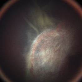
 Cohen Syndrome Retinal Detachment
Apr 30 2020 by Giselle DeOliveira
Gonio Photograph of 13-month infant male with retinal detachment, retinopathy of prematurity and Cohen Syndrome
Photographer: Giselle DeOliveira, University of Miami, Bascom Palmer Eye Institute
Imaging device: Retcam III
Condition/keywords: retinopathy of prematurity (ROP)
-
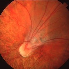
 Dragged Disc from Retinopathy of Prematurity
Apr 2 2019 by Gary R. Cook, MD, FACS
36-year-old white fenale with dragged disc OS secondary to cicatricial retinopathy of prematurity; V.A. = 20/80
Imaging device: Topcon VT-50
Condition/keywords: cicatricial retinopathy of prematurity, dragged disc, retinopathy of prematurity (ROP)
-
 Fibrotic Tractional Membrane in ROP Stage 5
Nov 7 2013 by Maria Ana Martinez-Castellanos, MD
Stage 5 retinopathy of prematurity in a 6 month old baby.
Photographer: Maria A. Martinez-Castellanos. Asociacion para Evitar la Ceguera en Mexico
Imaging device: RetCam II
Condition/keywords: fibrous proliferation, fibrovascular proliferation, retinopathy of prematurity (ROP)
-
 Fight for Sight
Mar 26 2024 by Tushar Agrawal
Fundus photograph showing 28 weeker APROP; regressed well after ROP Laser photocoagulation as seen at age 3 months.
Imaging device: Retcam neo
Condition/keywords: aggressive posterior retinopathy of prematurity (APROP), pediatric retina, retinopathy of prematurity (ROP)
-
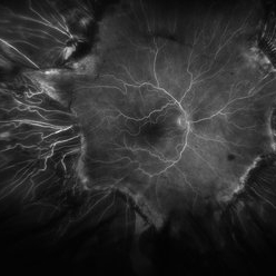 Fluorescein Angiogram of ROP With Cryo Scarring
Jul 7 2025 by Jenn Geelan
FA photo of a 34 year old male with prior stage 3 ROP with history of 360 degree cryotherapy.
Photographer: Jenn Geelan, Retina-Vitreous Surgeons of CNY
Imaging device: Optos California
Condition/keywords: cryotheraphy scar, fluorescein angiogram (FA), fundus photograph, retinopathy of prematurity (ROP), ROP, tilted disc
-
 Flying Baby Imaging Technique
May 27 2020 by Jamin S. Brown, MD
7-month-old male with improving ROP.
Photographer: Stefanie Palmer CRA, Retina-Vitreous Surgeons of CNY
Condition/keywords: retinopathy of prematurity (ROP)
-
 Fundus with ROP OD
Feb 4 2021 by Abdallah Mahmoud
Fundus with retinopathy or prematurity OD.
Condition/keywords: fundus photograph, OD, retinopathy of prematurity (ROP)
-
 Fundus with ROP OS
Feb 4 2021 by Abdallah Mahmoud
Fundus with retinopathy of prematurity OS
Condition/keywords: fundus photograph, retinopathy of prematurity (ROP)
-
 Iridocorneal Angle
Sep 13 2013 by Maria Ana Martinez-Castellanos, MD
Angle OCT of a right eye of a 4 months old male patient with retinopathy of prematurity stage 5.
Photographer: Maria A. Martinez-Castellanos. Asociación para Evitar la Ceguera en Mexico
Condition/keywords: angle closure, retinopathy of prematurity (ROP), retinopathy of prematurity, stage 5
-
 Latrogenic Oxygen Induce Retinopathy in a Premature Baby
Mar 13 2013 by Maria Ana Martinez-Castellanos, MD
Angiogram taken on a 42 weeks corrected age baby born at 34 weeks. The baby developed lung disease and received oxygen 100% for 4 weeks.
Photographer: Maria A. Martinez-Castellanos. Asociacion para Evitar la Ceguera en Mexico
Imaging device: RetCam II
Condition/keywords: retinopathy of prematurity (ROP)
-
 Lobular Choroidal Filling in ROP Stage 1
Nov 3 2013 by Maria Ana Martinez-Castellanos, MD
Lobular choroidal filling in a baby with stage 1 ROP, the macular area has a different filling pattern that the rest of the retina.
Photographer: Maria A. Martinez-Castellanos. Asociacion para Evitar la Ceguera en Mexico
Imaging device: RetCam II
Condition/keywords: choriocapillaris, retinopathy of prematurity (ROP)
-
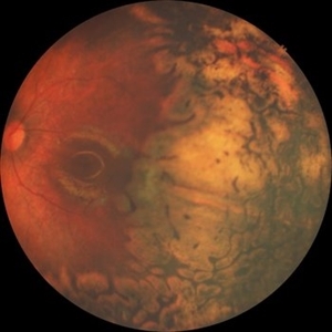
 Macula Sparring Tractional Retinal Detachment
Feb 9 2018 by Olivia Rainey
Ultra-wide field pseudocolor image of a 22-year-old male with a macula sparring tractional retinal detachment relating to retinopathy of prematuritiy affecting his right eye.
Photographer: Olivia Rainey
Imaging device: Optos
Condition/keywords: color fundus photograph, demarcation line, macula sparring, Optos, retinopathy of prematurity (ROP), tractional retinal detachment, ultra-wide field imaging
-
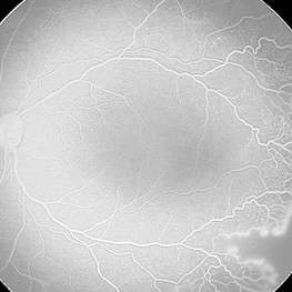
 Plus Disease
Aug 20 2018 by Anna L. Ells, MD, FRCS(C)
Wide field image of right eye with severe ROP with Plus disease.
Photographer: Anna Ells, Calgary Retina Consultants
Imaging device: RetCam
Condition/keywords: retinopathy of prematurity (ROP)
-
 Retinal Detachment
Apr 30 2020 by Giselle DeOliveira
Fundus Photograph of 13-month old male infant with retinopathy of prematurity, retinal detachment and Cohen Syndrome.
Photographer: Giselle DeOliveira, University of Miami, Bascom Palmer Eye Institute
Imaging device: Retcam III
Condition/keywords: retinopathy of prematurity (ROP)
-
 Retinal Detachment post ROP
Jul 22 2021 by Vishal Gupta, MBBS, MS
Fundus image of total Retinal Detachment in a five-year-old male kid with a history of prematurity.
Photographer: Dr Shobhit Chawla, Prakash Netra Kendr, Lucknow, UP, INDIA
Imaging device: Zeiss Clarus
Condition/keywords: retinopathy of prematurity (ROP)
-
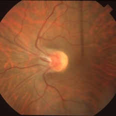
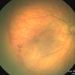
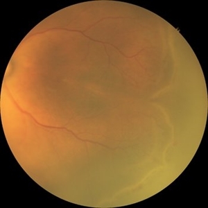 Retinopathy of Prematurity
Oct 26 2025 by Anjana Mirajkar, MS Ophthalmology
Fundus photograph of left eye premature baby having stage 3 in zone 2A with a secondary notch.
Photographer: Dr. Anjana Mirajkar- HV Desai eye hospital ,Pune
Imaging device: retcam
Condition/keywords: retinopathy of prematurity (ROP), stage 3
-
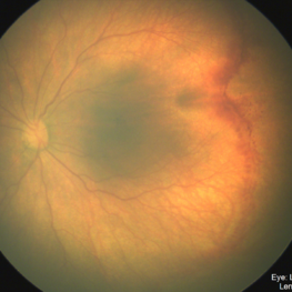
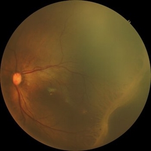 Retinopathy of Prematurity
Oct 26 2025 by Anjana Mirajkar, MS Ophthalmology
Fundus photograph of a left eye of a premature baby showing stage 3 in zone 2 posterior.
Photographer: Dr. Anjana Mirajkar- HV desai eye hospital ,Pune
Imaging device: Retcam
Condition/keywords: retinopathy of prematurity (ROP), retinopathy of prematurity stage 3
 Loading…