Search results (130 results)
-
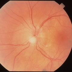
 Chorioretinal Scar
Feb 19 2024 by Sanauddin Samejo , Diploma (Ophthalmic Technician Training Course)
A patient came in to the clinic of Dr Madhav Rao (VR Surgeon).
Photographer: Sanauddin Samejo, Burjeel Hospital, Abu Dhabi, UAE.
Imaging device: Optos Silverstone
Condition/keywords: retinal scar
-
 Extensive/ Heavy Focal/ PRP OS - FAF
Jun 28 2018 by Hosam Attia, MD
70-year-old woman, seen for initial eye exam, with endstage PDR and H/O prior Focal/ PRP OU , somewhere else.
Imaging device: Optos - California
Condition/keywords: chorioretinal scar, ghost vessels, laser scarring, optic atrophy, pan-retinal photocoagulation (PRP), proliferative diabetic retinopathy (PDR), retinal scar
-
---thumb.jpg/image-square;max$300,300.ImageHandler) Retinal Scar
Retinal Scar
Feb 13 2013 by From the Collections of Thomas M. Aaberg, MD and Thomas M. Aaberg Jr., MD
Retinal scar.
Condition/keywords: retinal scar
-
 Acute Toxoplasmosis in AIDS
Apr 8 2019 by Gary R. Cook, MD, FACS
Left eye of a white male with AIDS and an optic neuritis secondary to ocular toxoplasmosis infection. The patient had no pre-existing chorioretinal scars secondary to Toxo. An edematous optic nerve with a focus of active retinitis inferonasally, small surface hemorrhage above it, and surrounding peripapillary edema is visible.
Imaging device: Topcon VT-50
Condition/keywords: AIDS, ocular toxoplasmosis, optic neuritis, toxoplasmosis
-
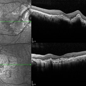 Angioid Streak-Associated Choroidal Neovasclar Membranes
Dec 27 2016 by Young Hee Yoon, MD, PhD
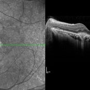
Optical coherence tomogaphs of an 74-year-old woman who received several anti-VEGF injections due to CNV associated with angioid streak in both eyes. There are diffuse CNVM in her right eye and subretinal scar in her left eye. Note the irregular crack in IR image of right eye and the focal Bruch's membrane dehiscence in corresponding B-scan image.
Photographer: Young Hee Yoon, University of Ulsan, Asan Medical Center, Seoul, Korea
Imaging device: Spectralis
Condition/keywords: angioid streaks, choroidal neovascularization (CNV)
-
 ARMD
ARMD
Aug 7 2013 by H. Michael Lambert, MD
Marked subretinal scarring in AMD.
Condition/keywords: central disciform scar, subretinal bands
-
---thumb.jpg/image-square;max$300,300.ImageHandler) case 2 OD
case 2 OD
Feb 14 2013 by From the Collections of Thomas M. Aaberg, MD and Thomas M. Aaberg Jr., MD
reproductions of figures 4 and 5 from the article "Ocular involvement in neonatal herpes simplex virus infection" (Hagler WS et al, Arch Opthalmol 1969;82:169-76.). Fulminating chorioretinal scarring and retinal pigmentary changes were seen in both eyes of an infant with neonatal systemic herpesvirus infection.
Condition/keywords: chorioretinal scar, neonatal herpes
-
 Central Retinal Vein Occlusion
Jul 13 2018 by Olivia Rainey
Ultra-wide field, pseudocolor montage of a patient presenting with a central retinal vein occlusion, as well as, an inferior chorioretinal scar in their right eye.
Photographer: Olivia Rainey
Imaging device: Optos
Condition/keywords: central retinal vein occlusion (CRVO), chorioretinal scar, montage, Optos, pseudocolor, ultra-wide field imaging
-
 Chorio Retinal Scar
Feb 19 2024 by Sanauddin Samejo , Diploma (Ophthalmic Technician Training Course)
A patient came in to Clinic of Dr Madhav Rao (VR Surgeon)
Photographer: Sanauddin Samejo, Burjeel Hospital, Abu Dhabi, UAE.
Imaging device: Optos Silverstone
Condition/keywords: chorioretinal scar
-
 Chorioretinal Macula Scar (Macula View)
May 12 2025 by Briana Hernandez
Zoomed in Macular View of Chorioretinal Macular Scar in 9-year-old female patient.
Photographer: Briana Hernandez, Hilton Head Retina Insitute
Imaging device: Optos
Condition/keywords: chorioretinal scar
-
 Chorioretinal Macula Scar (Ultrawide View)
May 12 2025 by Briana Hernandez
Ultra wide Optos image of Chorioretinal Macular Scar in 9-year-old female patient.
Photographer: Briana Hernandez, Hilton Head Retina Institute
Imaging device: Optos
Condition/keywords: chorioretinal scar, macular scar, ultra-wide field imaging
-
 Chorioretinal Scar
Apr 1 2016 by Nichole Lewis
Chorioretinal scar.
Photographer: Nichole Lewis - Pennsylvania Retina Specialists, Camp Hill, PA
Condition/keywords: chorioretinal scar
-
 Chorioretinal Scar
May 16 2017 by Olivia Rainey
Fundus photograph of an 17-year-old male with a macular scar affecting his right eye secondary to exudation from Coats disease.
Photographer: Olivia Rainey
Imaging device: Topcon 50dx
Condition/keywords: 20 degrees, chorioretinal scar, Coats' disease, color fundus photograph, color photo, fundus photograph
-
---thumb.jpg/image-square;max$300,300.ImageHandler) Chorioretinal Scarring
Chorioretinal Scarring
Feb 15 2013 by From the Collections of Thomas M. Aaberg, MD and Thomas M. Aaberg Jr., MD
Color fundus photograph showing chorioretinal scarring consistent with prior retinal laser photocoagulation to areas of peripheral retinal nonperfusion.
Condition/keywords: laser scarring, peripheral retinal nonperfusion
-
 Chorioretinal Scars with Subretinal Fibrosis and an old Retinal Detachment
May 3 2018 by Nichole Lewis
Chorioretinal scars with subretinal fibrosis and an old retinal detachment.
Photographer: Nichole Lewis
Condition/keywords: chorioretinal scar, chronic retinal detachment, subretinal fibrosis
-
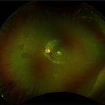
 Chorioretinitis with Overlying Vitreous Stranding/Vitritis
Mar 23 2023 by Isaac Agranoff
Fundus photograph of a 37-year-old woman presenting with chorioretinitis with overlying vitreous stranding/vitritis that has remained unchanged for multiple years. Patient presented with irritation and blurred vision and her vision was 20/40 OD. The OCT revealed evidence of low-grade inflammation and the recommend treatment was anti-inflammatory eye drops at this time and to obtain second opinion with another physician in the office.
Photographer: Isaac Agranoff, Technician
Imaging device: Optos California
Condition/keywords: chorioretinal scar, chorioretinitis, inflammation, Optos, ultra-wide field imaging, vitritis
-
 Choroidal Hemangioma
Oct 20 2012 by Hyung-Woo Kwak, MD
Fundus, ICG, and OCT examination showed a typical chorioretinal scar lying concentric to the optic disc. Typical choroidal rupture was performed after intravitreal gas injection under the diagnosis of submacular hemorrhage caused by trauma, after the absorption of submacular hemorrhage
Condition/keywords: chorioretinal scar, choroidal rupture, submacular hemorrhage
-
 Choroidal Hemangioma
Oct 20 2012 by Hyung-Woo Kwak, MD
Fundus, ICG and OCT examination showed a typical chorioretinal scar lying concentric to the optic disc. Typical choroidal rupture was performed after intravitreal gas injection under the diagnosis of submacular hemorrhage caused by trauma, after the absorption of submacular hemorrhage
-
 Choroidopathy
May 27 2020 by Jamin S. Brown, MD
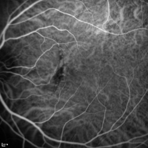
Fluorescein angiography image of 28 year old female with focal chorioretinal inflammation, macular or paramacular OS. chorioretinal scar OS.
Photographer: Jeffrey Barker, Retina-Vitreous Surgeons of CNY
Condition/keywords: choroidopathy
-
 CNV Due to Toxoplasmosis
Apr 6 2014 by Ratimir Lazic, MD, PhD
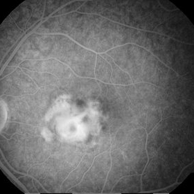
A FAG image of a 7-year-old boy. Late venous phase image shows unsharply limited leakage of dye which presents staining of chorioretinal scar with active CNV.
Photographer: Marko Vlasic, University Eye Clinic Svjetlost
Imaging device: Zeis Visucam Lite 2
Condition/keywords: choroidal neovascularization (CNV), toxoplasmosis
-
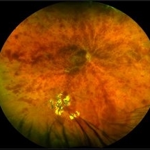
 CNV due to Toxoplasmosis
Apr 6 2014 by Ratimir Lazic, MD, PhD
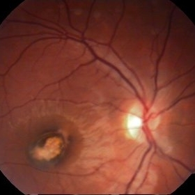
A color fundus image of a 7-year-old boy. Pigmented chorioretinal scar sorrounded by subretinal hemorrhage can be seen. VA is 0,2 by Snellen lines. The image presents the baseline clinical picture. The antiVEGF intravitreal injection, under general anesthesia, was administered.
Photographer: Marko Vlasic, University Eye Clinic Svjetlost
Imaging device: Zeis Visucam Lite 2
Condition/keywords: choroidal neovascularization (CNV), subretinal hemorrhage, toxoplasmosis
-
---thumb.JPG/image-square;max$300,300.ImageHandler) Concussion With Choroidal Rupture
Concussion With Choroidal Rupture
Dec 13 2013 by Mallika Goyal, MD
Fundus photograph of a 26-year-old male 4 weeks following head injury in a road accident. VA is 20/20 but quality is subnormal; there is afferent pupillary defect, choroidal rupture passing through foveal centre, likely traumatic optic neuropathy (in view of RAPD), submacular heme and extensive chorioretinal scarring inferior to macula, vitreous haemorrhage.
Photographer: Mallika Goyal, MD, Apollo Health City, Hyderabad, India
Condition/keywords: choroidal rupture, concussion
-
---thumb.JPG/image-square;max$300,300.ImageHandler) Concussion With Choroidal Rupture
Concussion With Choroidal Rupture
Dec 13 2013 by Mallika Goyal, MD
Fundus photograph of a 26-year-old male 4 weeks following head injury in a road accident. VA is 20/20 but quality is subnormal; there is afferent pupillary defect, choroidal rupture passing through foveal centre, likely traumatic optic neuropathy (in view of RAPD), submacular heme and extensive chorioretinal scarring inferior to macula, vitreous hemorrhage.
Photographer: Mallika Goyal, MD, Apollo Health City, Hyderabad, India
Condition/keywords: choroidal rupture, concussion
 Loading…