Search results (77 results)
-
 ARMD / CNVM / PED With RPE Tear
Nov 7 2014 by David Callanan, MD
59-year-old male, ARMD / CNVM / PED with RPE Tear.
Condition/keywords: choroidal neovascular membrane (CNVM), pigment epithelial detachment (PED), retinal pigment epithelium (RPE) tear
-
 ARMD / CNVM / PED With RPE Tear
Nov 7 2014 by David Callanan, MD
59-year-old male, ARMD / CNVM / PED with RPE Tear.
Condition/keywords: choroidal neovascular membrane (CNVM), pigment epithelial detachment (PED), retinal pigment epithelium (RPE) tear
-
 ARMD / CNVM / PED With RPE Tear
Nov 7 2014 by David Callanan, MD
59-year-old male, ARMD / CNVM / PED with RPE Tear.
Condition/keywords: choroidal neovascular membrane (CNVM), pigment epithelial detachment (PED), retinal pigment epithelium (RPE) tear
-
 ARMD / CNVM / PED With RPE Tear
Nov 7 2014 by David Callanan, MD
59-year-old male, ARMD / CNVM / PED with RPE Tear.
Condition/keywords: choroidal neovascular membrane (CNVM), pigment epithelial detachment (PED), retinal pigment epithelium (RPE) tear
-
 ARMD / CNVM / PED With RPE Tear
Nov 7 2014 by David Callanan, MD
59-year-old male, ARMD / CNVM / PED with RPE Tear.
Condition/keywords: choroidal neovascular membrane (CNVM), pigment epithelial detachment (PED), retinal pigment epithelium (RPE) tear
-
 ARMD with RPE Rip
Oct 12 2012 by Jeffrey G. Gross, MD, FASRS
ARMD with RPE rip.
Condition/keywords: retinal pigment epithelium, retinal pigment epithelium (RPE) tear
-
 ARMD with RPE Rip
Oct 12 2012 by Jeffrey G. Gross, MD, FASRS
ARMD with RPE rip, FA, showing window defect and blockage from retracted RPE layer.
Condition/keywords: retinal pigment epithelium, retinal pigment epithelium (RPE) tear, retracted retinal pigment epithelium (RPE) layer
-
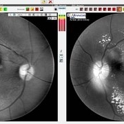
 Extrafoveal PED with RPE rip AF
Dec 23 2012 by Alex P. Hunyor, MD
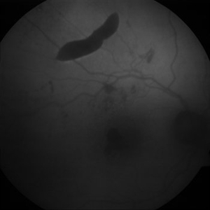
80-year-old female with subfoveal occult CNV and large extrafoveal PED which underwent spontaneous RPE rip. Autofluorescence image shows hypoautofluorescence in crescentic area of absent RPE due to rip, and also RPE atrophy adjacent to fovea. Intervening small areas of hypoautofluorescence are due to subretinal haemorrhage.
Condition/keywords: pigment epithelial detachment (PED), retinal pigment epithelium (RPE) tear
-
 Extrafoveal PED with RPE rip colour photo
Dec 23 2012 by Alex P. Hunyor, MD
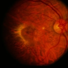
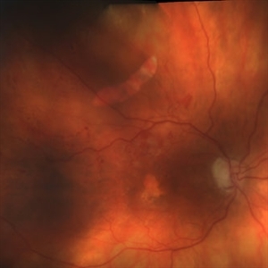
80-year-old female with subfoveal occult CNV and large extrafoveal PED which underwent spontaneous RPE rip.
Condition/keywords: pigment epithelial detachment (PED), retinal pigment epithelium (RPE) tear
-
 Extrafoveal PED with RPE rip FA1
Dec 23 2012 by Alex P. Hunyor, MD
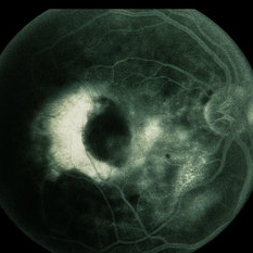
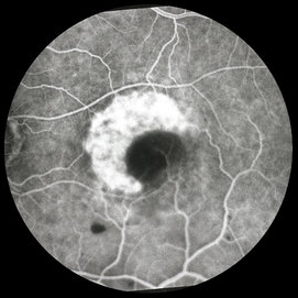
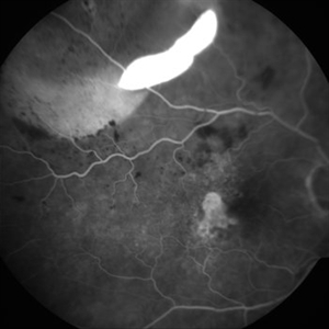
80-year-old female with subfoveal occult CNV and large extrafoveal PED which underwent spontaneous RPE rip. Early phase FA showing intense hyperfluorescence in the area of acute absence of RPE.
Condition/keywords: pigment epithelial detachment (PED), retinal pigment epithelium (RPE) tear
-
 Extrafoveal PED with RPE rip FA2
Dec 23 2012 by Alex P. Hunyor, MD
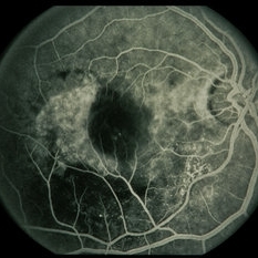
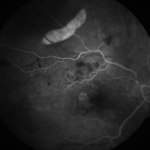
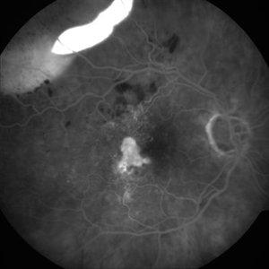
80-year-old female with subfoveal occult CNV and large extrafoveal PED which underwent spontaneous RPE rip. FA shows intense hyperfluorescence in area of absent RPE, progressive filling of extrafoveal PED, and hyperfluorescence in macula from atrophy and occult CNV.
Condition/keywords: pigment epithelial detachment (PED), retinal pigment epithelium (RPE) tear
-
 Extrafoveal PED with RPE rip FA3
Dec 23 2012 by Alex P. Hunyor, MD
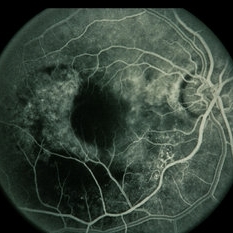
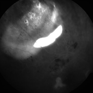
80-year-old female with subfoveal occult CNV and large extrafoveal PED which underwent spontaneous RPE rip. FA shows intense hyperfluorescence in area of absent RPE, progressive filling of extrafoveal PED, and hyperfluorescence in macula from atrophy and occult CNV.
Condition/keywords: pigment epithelial detachment (PED), retinal pigment epithelium (RPE) tear
-
 Extrafoveal PED with RPE rip FA4
Dec 23 2012 by Alex P. Hunyor, MD
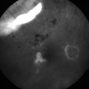
80-year-old female with subfoveal occult CNV and large extrafoveal PED which underwent spontaneous RPE rip. FA shows intense hyperfluorescence in area of absent RPE, progressive filling of extrafoveal PED, and hyperfluorescence in macula from atrophy and occult CNV.
Condition/keywords: pigment epithelial detachment (PED), retinal pigment epithelium (RPE) tear
-
 Extrafoveal PED with RPE rip FA5
Dec 23 2012 by Alex P. Hunyor, MD
80-year-old female with subfoveal occult CNV and large extrafoveal PED which underwent spontaneous RPE rip. FA shows intense hyperfluorescence in area of absent RPE, progressive filling of extrafoveal PED, and hyperfluorescence in macula from atrophy and occult CNV.
Condition/keywords: pigment epithelial detachment (PED), retinal pigment epithelium (RPE) tear
-
 RIP 1 FAF
Oct 7 2015 by Roberto Gallego-Pinazo, MD, PhD, DiSSO
Multicolor and autofluorescence sequence of a retinal pigment epithelium tear following intravitreal anti-VEGF injection.
Photographer: Rosa Dolz-Marco, University and Polytechnic Hospital La Fe, Valencia, Spain
Condition/keywords: age-related macular degeneration (AMD), autofluorescence imaging, choroidal neovascularization (CNV), multicolor, retinal pigment epithelium (RPE) tear
-
 RIP 2 FAF
Oct 7 2015 by Roberto Gallego-Pinazo, MD, PhD, DiSSO
Multicolor and autofluorescence sequence of a retinal pigment epithelium tear following intravitreal anti-VEGF injection.
Photographer: Rosa Dolz-Marco, University and Polytechnic Hospital La Fe, Valencia, Spain
Condition/keywords: age-related macular degeneration (AMD), autofluorescence imaging, choroidal neovascularization (CNV), multicolor, retinal pigment epithelium (RPE) tear
-
 RIP1
RIP1
Oct 7 2015 by Roberto Gallego-Pinazo, MD, PhD, DiSSO
Multicolor and autofluorescence sequence of a retinal pigment epithelium tear following intravitreal anti-VEGF injection.
Photographer: Rosa Dolz-Marco, University and Polytechnic Hospital La Fe, Valencia, Spain
Condition/keywords: age-related macular degeneration (AMD), autofluorescence imaging, choroidal neovascularization (CNV), multicolor, retinal pigment epithelium (RPE) tear
-
 RIP2
RIP2
Oct 7 2015 by Roberto Gallego-Pinazo, MD, PhD, DiSSO
Multicolor and autofluorescence sequence of a retinal pigment epithelium tear following intravitreal anti-VEGF injection.
Photographer: Rosa Dolz-Marco, University and Polytechnic Hospital La Fe, Valencia, Spain
Condition/keywords: age-related macular degeneration (AMD), autofluorescence imaging, choroidal neovascularization (CNV), multicolor, retinal pigment epithelium (RPE) tear
-
 RPE Micro Rip in Central Serous Chorioretinopathy
Jun 26 2016 by Rameez N Hussain, MD
SD OCT image of a case of central serous retinopathy showing RPE micro rip (RPE leak).
Photographer: DR RAMEEZ N HUSSAIN
Imaging device: Dense scan mode - Heidelberg Spectralis
Condition/keywords: central serous chorioretinopathy (CSCR), focal laser, leakage, retinal pigment epithelium (RPE) tear, Spectralis
-
 RPE Rip
Feb 13 2013 by From the Collections of Thomas M. Aaberg, MD and Thomas M. Aaberg Jr., MD
RPE rip, foveal CNV
Condition/keywords: choroidal neovascularization (CNV), macular laser, retinal pigment epithelium (RPE) tear
-
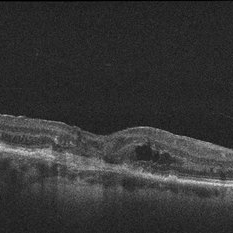 RPE rip macular OCT
Dec 23 2012 by Alex P. Hunyor, MD
80-year-old female with subfoveal occult CNV and large extrafoveal PED which underwent spontaneous RPE rip. OCT shows subfoveal CNV and intraretinal cystic edema
Condition/keywords: pigment epithelial detachment (PED), retinal pigment epithelium (RPE) tear
-
 RPE Tear
Apr 21 2014 by Xiaoxin Li, MD PhD
Multispectral digital ophthalmoscope of a 64-year-old woman with a RPE tear. (red-green)
Photographer: Xiaoxin Li, Xinxin Wang
Imaging device: multispectral fundus photography
Condition/keywords: retinal pigment epithelium (RPE) tear
-
 RPE Tear
Apr 21 2014 by Xiaoxin Li, MD PhD
Multispectral digital ophthalmoscope of a 64-year-old woman with a RPE tear. (590 nm)
Photographer: Xiaoxin Li, Xinxin Wang
Imaging device: multispectral fundus photography
Condition/keywords: retinal pigment epithelium (RPE) tear
-
 RPE Tear
Apr 21 2014 by Xiaoxin Li, MD PhD
Multispectral digital ophthalmoscope of a 64-year-old woman with a RPE tear. (660 nm)
Photographer: Xiaoxin Li, Xinxin Wang
Imaging device: multispectral fundus photography
Condition/keywords: retinal pigment epithelium (RPE) tear
-
 RPE Tear
Apr 21 2014 by Xiaoxin Li, MD PhD
Multispectral digital ophthalmoscope of a 64-year-old woman with a RPE tear. (760 nm)
Photographer: Xiaoxin Li, Xinxin Wang
Imaging device: multispectral fundus photography
Condition/keywords: retinal pigment epithelium (RPE) tear
 Loading…