Search results (33 results)
-
---thumb.jpg/image-square;max$300,300.ImageHandler) Central Serous Chorioretinopathy 1
Central Serous Chorioretinopathy 1
Mar 18 2013 by Maurice F. Rabb
Woman with a 3 month history of reduced vision, and her fundi appeared as if she had a severe form of central serous chorioretinopathy, including subretinal febrin deposition, serous pigment epithelial detachments, patchy zones of pigment epithelial atrophy, and dependent, bullous detachments bilaterally. There are also multifocal areas of orange subretinal deposits, some in the form of an irregular sequence or change. These looked like Elschnig spots and Siegrist lines, consistent with choroidal ischemia that could account for the exudative detachments as well.
Condition/keywords: bullous detachments bilaterally, central serous chorioretinopathy (CSCR), choroidal ischemia, dependent, orange subretinal deposits, patchy zones of pigment epithelial atrophy, reduced vision, serous pigment epithelial detachment, Siegrist Streaks, subretinal fibrin deposition
-
---thumb.jpg/image-square;max$300,300.ImageHandler) Central Serous Chorioretinopathy 2
Central Serous Chorioretinopathy 2
Mar 18 2013 by Maurice F. Rabb
Woman with a 3 month history of reduced vision, and her fundi appeared as if she had a severe form of central serous chorioretinopathy, including subretinal febrin deposition, serous pigment epithelial detachments, patchy zones of pigment epithelial atrophy, and dependent, bullous detachments bilaterally. There are also multifocal areas of orange subretinal deposits, some in the form of an irregular sequence or change. These looked like Elschnig spots and Siegrist lines, consistent with choroidal ischemia that could account for the exudative detachments as well.
Condition/keywords: bullous detachments bilaterally, central serous chorioretinopathy (CSCR), choroidal ischemia, dependent, orange subretinal deposits, patchy zones of pigment epithelial atrophy, reduced vision, serous pigment epithelial detachment, Siegrist Streaks, subretinal fibrin deposition
-
---thumb.jpg/image-square;max$300,300.ImageHandler) Central Serous Chorioretinopathy 3
Central Serous Chorioretinopathy 3
Mar 18 2013 by Maurice F. Rabb
Woman with a 3 month history of reduced vision, and her fundi appeared as if she had a severe form of central serous chorioretinopathy, including subretinal febrin deposition, serous pigment epithelial detachments, patchy zones of pigment epithelial atrophy, and dependent, bullous detachments bilaterally. There are also multifocal areas of orange subretinal deposits, some in the form of an irregular sequence or change. These looked like Elschnig spots and Siegrist lines, consistent with choroidal ischemia that could account for the exudative detachments as well.
Condition/keywords: bullous detachments bilaterally, central serous chorioretinopathy (CSCR), choroidal ischemia, dependent, orange subretinal deposits, patchy zones of pigment epithelial atrophy, reduced vision, serous pigment epithelial detachment, subretinal fibrin deposition
-
---thumb.jpg/image-square;max$300,300.ImageHandler) Central Serous Chorioretinopathy 4
Central Serous Chorioretinopathy 4
Mar 18 2013 by Maurice F. Rabb
Woman with a 3 month history of reduced vision, and her fundi appeared as if she had a severe form of central serous chorioretinopathy, including subretinal febrin deposition, serous pigment epithelial detachments, patchy zones of pigment epithelial atrophy, and dependent, bullous detachments bilaterally. There are also multifocal areas of orange subretinal deposits, some in the form of an irregular sequence or change. These looked like Elschnig spots and Siegrist lines, consistent with choroidal ischemia that could account for the exudative detachments as well.
Condition/keywords: bullous detachments bilaterally, central serous chorioretinopathy (CSCR), choroidal ischemia, dependent, orange subretinal deposits, patchy zones of pigment epithelial atrophy, reduced vision, serous pigment epithelial detachment, Siegrist Streaks, subretinal fibrin deposition
-
---thumb.jpg/image-square;max$300,300.ImageHandler) Central Serous Chorioretinopathy 5
Central Serous Chorioretinopathy 5
Mar 18 2013 by Maurice F. Rabb
Woman with a 3 month history of reduced vision, and her fundi appeared as if she had a severe form of central serous chorioretinopathy, including subretinal febrin deposition, serous pigment epithelial detachments, patchy zones of pigment epithelial atrophy, and dependent, bullous detachments bilaterally. There are also multifocal areas of orange subretinal deposits, some in the form of an irregular sequence or change. These looked like Elschnig spots and Siegrist lines, consistent with choroidal ischemia that could account for the exudative detachments as well.
Condition/keywords: bullous detachments bilaterally, central serous chorioretinopathy (CSCR), dependent, orange subretinal deposits, patchy zones of pigment epithelial atrophy, reduced vision, serous pigment epithelial detachment, Siegrist Streaks, subretinal fibrin deposition
-
---thumb.jpg/image-square;max$300,300.ImageHandler) Central Serous Chorioretinopathy 6
Central Serous Chorioretinopathy 6
Mar 18 2013 by Maurice F. Rabb
Woman with a 3 month history of reduced vision, and her fundi appeared as if she had a severe form of central serous chorioretinopathy, including subretinal febrin deposition, serous pigment epithelial detachments, patchy zones of pigment epithelial atrophy, and dependent, bullous detachments bilaterally. There are also multifocal areas of orange subretinal deposits, some in the form of an irregular sequence or change. These looked like Elschnig spots and Siegrist lines, consistent with choroidal ischemia that could account for the exudative detachments as well.
Condition/keywords: bullous detachments bilaterally, central serous chorioretinopathy (CSCR), choroidal ischemia, dependent, orange subretinal deposits, patchy zones of pigment epithelial atrophy, reduced vision, serous pigment epithelial detachment, Siegrist Streaks, subretinal fibrin deposition
-
---thumb.jpg/image-square;max$300,300.ImageHandler) Central Serous Chorioretinopathy 7
Central Serous Chorioretinopathy 7
Mar 18 2013 by Maurice F. Rabb
Woman with a 3 month history of reduced vision, and her fundi appeared as if she had a severe form of central serous chorioretinopathy, including subretinal febrin deposition, serous pigment epithelial detachments, patchy zones of pigment epithelial atrophy, and dependent, bullous detachments bilaterally. There are also multifocal areas of orange subretinal deposits, some in the form of an irregular sequence or change. These looked like Elschnig spots and Siegrist lines, consistent with choroidal ischemia that could account for the exudative detachments as well.
Condition/keywords: bullous detachments bilaterally, central serous chorioretinopathy (CSCR), choroidal ischemia, dependent, orange subretinal deposits, reduced vision, serous pigment epithelial detachment, Siegrist Streaks, subretinal fibrin deposition
-
---thumb.jpg/image-square;max$300,300.ImageHandler) Central Serous Chorioretinopathy 8
Central Serous Chorioretinopathy 8
Mar 18 2013 by Maurice F. Rabb
Woman with a 3 month history of reduced vision, and her fundi appeared as if she had a severe form of central serous chorioretinopathy, including subretinal febrin deposition, serous pigment epithelial detachments, patchy zones of pigment epithelial atrophy, and dependent, bullous detachments bilaterally. There are also multifocal areas of orange subretinal deposits, some in the form of an irregular sequence or change. These looked like Elschnig spots and Siegrist lines, consistent with choroidal ischemia that could account for the exudative detachments as well.
Condition/keywords: bullous detachments bilaterally, central serous chorioretinopathy (CSCR), choroidal ischemia, dependent, orange subretinal deposits, patchy zones of pigment epithelial atrophy, reduced vision, serous pigment epithelial detachment, Siegrist Streaks, subretinal fibrin deposition
-
---thumb.jpg/image-square;max$300,300.ImageHandler) Reduced Vision
Reduced Vision
Feb 4 2014 by Maurice F. Rabb
69-year-old female with a detachment of the pigment epithelium in the right eye with hemorrhagic and early fibrous proliferative changes. The left eye contained a large, turbid detachment of the pigment epithelium with patchy atrophy, a very shallow evident overlying sensory retinal detachment, but no subretinal hemorrhage. The visual acuity in each eye was 20/400.
Condition/keywords: fibrous proliferation, patchy atrophy, pigment epithelial detachment (PED), reduced vision
-
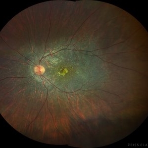 Bietti's Crystalline Dystrophy
Oct 3 2020 by SHISHIR VERGHESE, MS, FVRS, FAICO (Retina)
Fundus photo of the left eye of a 21-year-old gentleman with complaints of reduced vision, nyctalopia and visual field reduction. Fundus photograph showing numerous small glistening yellow-white retinal crystalline deposits in the retina.
Photographer: Shishir Verghese, Aravind Eye Hospital, Coimbatore
Condition/keywords: Bietti's crystalline dystrophy, heredomacular degeneration, retinal degeneration
-
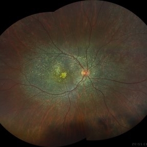 Bietti's Crystalline Dystrophy
Oct 3 2020 by SHISHIR VERGHESE, MS, FVRS, FAICO (Retina)
Fundus photo of the right eye of a 21-year-old gentleman with complaints of reduced vision, nyctalopia and visual field reduction. Fundus photograph showing numerous small glistening yellow-white retinal crystalline deposits in the retina.
Photographer: Shishir Verghese, Aravind Eye Hospital, Coimbatore
Condition/keywords: Bietti's crystalline dystrophy, hereditary retinal degeneration, heredomacular degeneration
 Loading…