Search results (300 results)
-
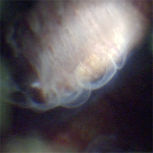
 Elevated Cystic Area
Nov 9 2012 by Norman Byer
This is the eye of a 53-year-old woman with a small elevated cystic area of the peripheral retina at the posterior end of a meridional fold.
Condition/keywords: meridional fold, ora serrata, peripheral retina, small elevated cystic area
-
---thumb.jpg/image-square;max$300,300.ImageHandler) Binder3 P12 Slide82
Binder3 P12 Slide82
Feb 15 2013 by From the Collections of Thomas M. Aaberg, MD and Thomas M. Aaberg Jr., MD
Color fundus photograph showing peripheral retinal nonperfusion, retinal neovascularization elsewhere (NVE), venous beading and dilatation, retinal microaneurysms, and intraretinal hemorrhage.
Condition/keywords: peripheral retinal nonperfusion, proliferative retinopathy, retinal neovascularization
-
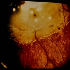
 Documented Retinal Pars Plana Cysts
Mar 14 2018 by Asaf Friehmann
Photograph taken during indentation of a 74-year-old patient who underwent a 25G pars plana vitrectomy (PPV) for repair of dislocated IOL, when this rarely documented peripheral retinal cyst which was found.
Photographer: Alexander Rubowitz
Condition/keywords: peripheral retinal cyst
-
---thumb.jpg/image-square;max$300,300.ImageHandler) Leakage from peripheral retinal neovascularization and peripheral nonperfusion
Leakage from peripheral retinal neovascularization and peripheral nonperfusion
Feb 15 2013 by From the Collections of Thomas M. Aaberg, MD and Thomas M. Aaberg Jr., MD
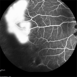
Late-phase fluorescein angiograph showing leakage from peripheral retinal neovascularization and peripheral nonperfusion
Condition/keywords: peripheral retinal nonperfusion, proliferative retinopathy, retinal neovascularization
-
 Peripheral Retinal Degeneration
Jul 8 2013 by Jason S. Calhoun
Patient in with double vision. VA was 20/20 in both eyes. Fundus exam shows retinal degenerative changes in both eyes. Offer to correct double vision with temporary Fresnel prism.
Photographer: Jason S. Calhoun, Department of Ophthalmology, Mayo Clinic Jacksonville, Florida
Condition/keywords: peripheral retinal degeneration
-
---thumb.JPG/image-square;max$300,300.ImageHandler) Peripheral Retinal Degeneration
Peripheral Retinal Degeneration
Jul 8 2013 by Jason S. Calhoun
Patient comes in with double vision. VA was 20/20 in both eyes. Fundus exam shows retinal degenerative changes in both eyes. Offer to correct double vision with temporary Fresnel prism.
Photographer: Jason S. Calhoun, Department of Ophthalmology, Mayo Clinic Jacksonville, Florida
Condition/keywords: peripheral retinal degeneration
-
 Peripheral Retinal Degeneration (L-ORD)
Apr 17 2024 by Virginia Gebhart
92 year old female with bilateral patchy, sharply demarcated circular areas of chorioretinal atrophy with hyperpigmented margins in the mid to far periphery. Labs showed normal plasma ornithine levels ruling out generalized gyrate atrophy. Also intermediate uveitis and CMD/CME. FTA-ABS, Quant gold, and HLA-A29 labs all negative.
Photographer: Virginia Gebhart
Imaging device: Optos California
Condition/keywords: cystoid macular degeneration, cystoid macular edema (CME), FA, Fluorescein angiography, peripheral retinal degeneration
-
 Peripheral retinal degenerations
Jan 29 2024 by Anupama Kiran Kumar
Fundus photo of a young man who underwent barrage laser after he presented to the clinic with floaters and was diagnosed to have lattices with horse shoe tears and retinal holes.
Photographer: Dr Anupama Kiran Kumar DNB FVR , Narayana Nethralaya Bangalore
Imaging device: Mirante SLO/OCT (Nidek Co., Gamagori, Japan)
Condition/keywords: lattice degeneration, peripheral retinal degeneration
-
---thumb.jpg/image-square;max$300,300.ImageHandler) Peripheral Retinal Dialysis & Subretinal Hemorrhage Cartoon
Peripheral Retinal Dialysis & Subretinal Hemorrhage Cartoon
Feb 13 2013 by From the Collections of Thomas M. Aaberg, MD and Thomas M. Aaberg Jr., MD
Peripheral retinal dialysis & subretinal hemorrhage cartoon.
Condition/keywords: cartoon, retinal hemorrhage, subretinal hemorrhage
-
Peripheral Retinal Hole with OCT Co-localization
Sep 26 2023 by Bradley T. Smith, MD, FASRS
Peripheral asymptomatic atrophic retinal hole with OCT co localization demonstrating small cuff of sub retinal fluid. Near infrared imaging shows hyper reflectivity through hole.
Condition/keywords: atrophic hole, lattice degeneration, OCT
-
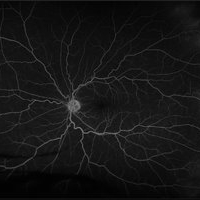 Peripheral Retinal Ischemia
Apr 26 2018 by Olivia Rainey
Ultra-wide field fluorescein angiogram of a 55-year-old female with peripheral retinal ischemia affecting her left eye. CTA head and neck performed on 11/16/15 and showed calcified atherosclerotic plaque involving the intracranial internal carotid arteries with resulting luminal narrowing. Intracranial vertebral arteries have smooth luminal contours. CTA neck normal. Likely from internal carotid plaques. Sickle cell disease came back negative.
Photographer: Olivia Rainey
Imaging device: Optos California
Condition/keywords: fluorescein angiogram (FA), fluorescein leakage, left eye, Optos, retinal ischemia, ultra-wide field imaging
-
 Peripheral Retinal Lesion
Nov 9 2012 by Norman Byer
This small elevated peripheral retinal lesion in a 48-year-old woman is a cystic retinal tuft. Such tufts are congenital developmental anomalies present from birth and situated behind the vitreous base. They are sites of abnormal vitreoretinal attachment, and can occasionally lead to retinal tears at the time of posterior vitreous detachment. They are present in about 5% of patients.
Condition/keywords: abnormal vitreal retinal attachment, behind the vitreous base, congenital anomaly, cystic retinal tuft, developmental anomaly, peripheral retinal lesion, present from birth
-
---thumb.jpg/image-square;max$300,300.ImageHandler) peripheral retinal nonperfusion, capillary abnormalities, leaking retinal microaneurysms, and blocked fluorescence
peripheral retinal nonperfusion, capillary abnormalities, leaking retinal microaneurysms, and blocked fluorescence
Feb 15 2013 by From the Collections of Thomas M. Aaberg, MD and Thomas M. Aaberg Jr., MD
Mid-phase fluorescein angiograph showing peripheral retinal nonperfusion, capillary abnormalities, leaking retinal microaneurysms, and blocked fluorescence from intraretinal hemorrhage.
Condition/keywords: peripheral retinal nonperfusion, proliferative retinopathy
-
---thumb.jpg/image-square;max$300,300.ImageHandler) Peripheral retinal nonperfusion, capillary abnormalities, retinal microaneurysms, and intraretinal hemorrhage
Peripheral retinal nonperfusion, capillary abnormalities, retinal microaneurysms, and intraretinal hemorrhage
Feb 15 2013 by From the Collections of Thomas M. Aaberg, MD and Thomas M. Aaberg Jr., MD
Color fundus photograph showing peripheral retinal nonperfusion, capillary abnormalities, retinal microaneurysms, and intraretinal hemorrhage.
Condition/keywords: peripheral retinal nonperfusion, proliferative retinopathy
-
---thumb.jpg/image-square;max$300,300.ImageHandler) Peripheral retinal nonperfusion, venous beading and dilatation, retinal microaneurysms, and intraretinal hemorrhage
Peripheral retinal nonperfusion, venous beading and dilatation, retinal microaneurysms, and intraretinal hemorrhage
Feb 15 2013 by From the Collections of Thomas M. Aaberg, MD and Thomas M. Aaberg Jr., MD
Color fundus photograph corresponding to slide titled "staining of retinal vessels, leakage from peripheral retinal neovascularization and peripheral nonperfusion." Shows peripheral retinal nonperfusion, venous beading and dilatation, retinal microaneurysms, and intraretinal hemorrhage.
Condition/keywords: peripheral retinal nonperfusion, proliferative retinopathy, retinal neovascularization
-
 Peripheral Retinal Tear
-
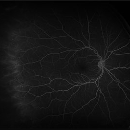 Peripheral Retinal Vasculitis
May 27 2020 by Olivia Rainey
Ultra-widefield fluorescein angiogram of a 58-year-old female with possible peripheral vasculitis. There was no venous access for this patient, so the fluorescein was administered orally. The image was taken at 7:33 after oral administration. The physician stated that the peripheral nonperfusion could be a sign of previous vasculitis, although could also be a result of uncontrolled diabetes. She was asked to obtain additional bloodwork in order to rule out sarcoidosis, as well as sickle cell. It does not appear the nonperfusion has progressed since her last evaluation. Her vision was 20/40 in the right eye at the time the image was taken.
Photographer: Olivia Rainey, OCT-C, COA
Imaging device: Optos California
Condition/keywords: diabetes, fluorescein angiogram (FA), hypertensive retinopathy, non-perfusion, Optos, oral fluorescein, peripheral retinal vasculitis, ultra-wide field imaging
-
 Pigmented Peripheral Retinal Degeneration
Jun 27 2013 by Jason S. Calhoun
42-year-old male came in for routine eye exam and to follow up on peripheral retinal degeneration in both eyes. VA is 20/20, right eye and 20/25, left eye. Patient is asymptomatic with no visual complaints.
Photographer: Jason S. Calhoun, Mayo Clinic Jacksonville, Florida
Imaging device: TOPCON TRC 50-EX
Condition/keywords: peripheral retinal degeneration
-
 Retinoblastoma
Dec 22 2014 by H. Michael Lambert, MD
Peripheral retinal photo of tumor
Condition/keywords: retinoblastoma, tumor
-
 Sickle Cell Retinopathy
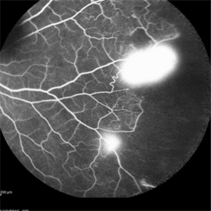
Sep 13 2015 by Thomas A. Ciulla, MD, MBA, FASRS
Angiography showed normal vessels posteriorly but severe capillary drop out throughout the periphery OU with scattered severe neovascularization at the edge of the capillary drop out peripherally.
Photographer: Thomas Steele
Condition/keywords: peripheral retinal neovascularization, sea fan, sickle cell retinopathy
-
 Sickle Cell Retinopathy
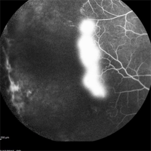
Sep 13 2015 by Thomas A. Ciulla, MD, MBA, FASRS
Angiography showed normal vessels posteriorly but severe capillary drop out throughout the periphery OU with scattered severe neovascularization at the edge of the capillary drop out peripherally.
Photographer: Thomas Steele
Condition/keywords: peripheral retinal neovascularization, sea fan, sickle cell retinopathy
-
 Sickle Cell Retinopathy
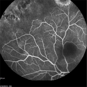
Sep 13 2015 by Thomas A. Ciulla, MD, MBA, FASRS
Angiography showed normal vessels posteriorly but severe capillary drop out throughout the periphery OU with scattered severe neovascularization at the edge of the capillary drop out peripherally.
Photographer: Thomas Steele
Condition/keywords: peripheral retinal neovascularization, sea fan, sickle cell retinopathy
-
 Sickle Cell Retinopathy
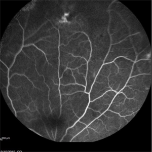
Sep 13 2015 by Thomas A. Ciulla, MD, MBA, FASRS
Angiography showed normal vessels posteriorly but severe capillary drop out throughout the periphery OU with scattered severe neovascularization at the edge of the capillary drop out peripherally.
Photographer: Thomas Steele
Condition/keywords: peripheral retinal neovascularization, sea fan, sickle cell retinopathy
-
 Sickle Cell Retinopathy
Sep 13 2015 by Thomas A. Ciulla, MD, MBA, FASRS
Angiography showed normal vessels posteriorly but severe capillary drop out throughout the periphery OU with scattered severe neovascularization at the edge of the capillary drop out peripherally.
Photographer: Thomas Steele
Condition/keywords: peripheral retinal neovascularization, sea fan, sickle cell retinopathy
 Loading…