Search results (45 results)
-
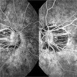 Fluorescein Angiography in High Myopia
Dec 7 2019 by Anfisa Ayalon, MD
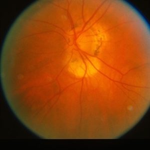
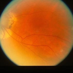
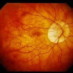
Fluorescein angiography pictures of a 55-year-old woman with high myopia.
Photographer: Anfisa Ayalon, MD., Meir Medical Center, Kfar Saba, Israel.
Condition/keywords: fluorescein angiogram (FA), high myopia, peripapillary atrophy
-
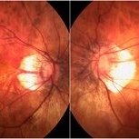 High Myopia
Dec 7 2019 by Anfisa Ayalon, MD
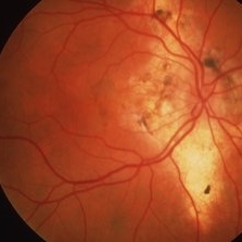
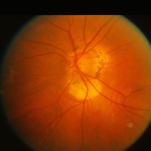
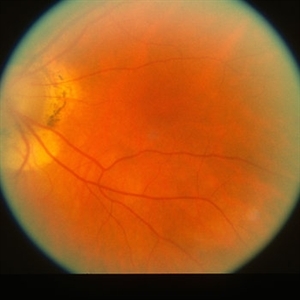
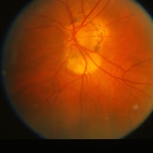
Fundus photograph of a 55-year-old woman with high myopia.
Photographer: Anfisa Ayalon,MD., Meir Medical Center, Kfar Saba, Israel.
Condition/keywords: high myopia, myopia, peripapillary atrophy
-
 Histo and Subfoveal Neovascular Membrane
Mar 27 2019 by Gary R. Cook, MD, FACS
41-year-old white female with a large subfoveal CNVM, subretinal fluid, and hemorrhage secondary to presumed ocular histoplasmosis (POHS) OS; V.A.= 20/400.
Imaging device: Topcon VT-50
Condition/keywords: hemorrhage, peripapillary atrophy, presumed ocular histoplasmosis syndrome (POHS), subfoveal choroidal neovascularization, subfoveal neovascular membrane
-
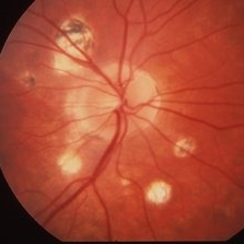 Histoplasmosis
Mar 27 2019 by Gary R. Cook, MD, FACS
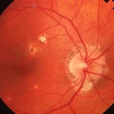
24-year-old white female with presumed ocular histoplasmosis (POHS) demonstrating some peripapillary atrophy and multiple atrophic histo spots around the optic nerve of her right eye; the patient was asymptomatic; V.A.= 20/20.
Imaging device: Topcon VT-50
Condition/keywords: atrophic spot, ocular histoplasmosis syndrome (OHS), peripapillary atrophy, presumed ocular histoplasmosis syndrome (POHS)
-
 Histoplasmosis and Old Disciform Macular Scar
Mar 27 2019 by Gary R. Cook, MD, FACS
Left eye of a 59-year-old white male with an old, inactive, disciform macular scar secondary to presumed ocular histoplasmosis (POHS); V.A.= counting fingers at 3 feet.
Imaging device: Topcon VT-50
Condition/keywords: central disciform scar, disciform scar, peripapillary atrophy, presumed ocular histoplasmosis syndrome (POHS)
-
 Histoplasmosis and Subfoveal Neovascular Membrane
Mar 27 2019 by Gary R. Cook, MD, FACS
Mid-phase (20.4 seconds) fluorescein angiogram image of the right eye of 59-year-old white male with ocular histoplasmosis and a well-defined subfoveal CNVM OD; V.A.= 20/80+2
Imaging device: Topcon VT-50
Condition/keywords: FA mid phase, fluorescein angiogram (FA), ocular histoplasmosis syndrome (OHS), peripapillary atrophy, presumed ocular histoplasmosis syndrome (POHS), subfoveal choroidal neovascularization, subfoveal neovascular membrane
-
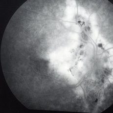 Histoplasmosis and Subfoveal Neovascular Membrane
Mar 27 2019 by Gary R. Cook, MD, FACS
Late-phase fluorescein angiogram image of the right eye of a 59-year-old white male with ocular histoplasmosis and a subfoveal neovascular membrane showing late leakage and diffusion of dye from the membrane; V.A.= 20/80+2.
Imaging device: Topcon VT-50
Condition/keywords: FA late phase, fluorescein angiogram (FA), ocular histoplasmosis syndrome (OHS), peripapillary atrophy, presumed ocular histoplasmosis syndrome (POHS), subfoveal neovascular membrane
-
 Histoplasmosis with Choroidal Neovascularization
Mar 27 2019 by Gary R. Cook, MD, FACS
59-year-old white male with presumed ocular histoplasmosis (POHS) and a choroidal neovascular membrane (CNVM) along the temporal margins of the peripapillary atrophy; V.A.= 20/80+2.
Imaging device: Topcon VT-50
Condition/keywords: choroidal neovascular membrane (CNVM), peripapillary atrophy, presumed ocular histoplasmosis syndrome (POHS)
-
 Ocular Histoplasmosis
Mar 27 2019 by Gary R. Cook, MD, FACS
Fellow eye (OD) of a 41-year-old white female with ocular histoplasmosis showing peripapillary atrophy and several atrophic histo spots OD; no CNVM present; V.A.= 20/20.
Imaging device: Topcon VT-50
Condition/keywords: atrophic spot, histoplasmosis, peripapillary atrophy, presumed ocular histoplasmosis syndrome (POHS)
-
 Peripapillary Atrophy
Oct 3 2014 by Mehul A Shah
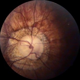
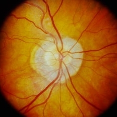
A 55-year-old patient presented with diminished vision OU on examination patient had glaucoma with peripapillary optic atrophy.
Photographer: Drashti Netralaya,Dahod
Imaging device: Zeiss ff450
Condition/keywords: atrophy, peripapillary
-
---thumb.jpg/image-square;max$300,300.ImageHandler) Peripapillary Atrophy
Peripapillary Atrophy
Feb 13 2013 by From the Collections of Thomas M. Aaberg, MD and Thomas M. Aaberg Jr., MD
Papilledema, intra-retinal hemorrhage, periopticneuritis.
Condition/keywords: intraretinal hemorrhage, papilledema, periopticneuritis, peripapillary atrophy
-
 Peripapillary Atrophy
Jul 31 2013 by From the Collections of Thomas M. Aaberg, MD and Thomas M. Aaberg Jr., MD
Peripapillary atrophy.
Condition/keywords: peripapillary atrophy
-
 Peripapillary Atrophy
Jul 31 2013 by From the Collections of Thomas M. Aaberg, MD and Thomas M. Aaberg Jr., MD
Peripapillary atrophy.
Condition/keywords: peripapillary atrophy
-
 Peripapillary Atrophy
Jul 31 2013 by From the Collections of Thomas M. Aaberg, MD and Thomas M. Aaberg Jr., MD
Peripapillary atrophy.
Condition/keywords: peripapillary atrophy
-
 Peripapillary Atrophy
Jul 31 2013 by From the Collections of Thomas M. Aaberg, MD and Thomas M. Aaberg Jr., MD
Peripapillary atrophy.
Condition/keywords: peripapillary atrophy
-
 Peripapillary Atrophy
Jul 31 2013 by From the Collections of Thomas M. Aaberg, MD and Thomas M. Aaberg Jr., MD
Peripapillary atrophy.
Condition/keywords: peripapillary atrophy
-
 Peripapillary Atrophy
Jul 31 2013 by From the Collections of Thomas M. Aaberg, MD and Thomas M. Aaberg Jr., MD
Peripapillary atrophy.
Condition/keywords: peripapillary atrophy
-
 Peripapillary Atrophy
Jul 31 2013 by From the Collections of Thomas M. Aaberg, MD and Thomas M. Aaberg Jr., MD
Peripapillary atrophy.
Condition/keywords: peripapillary atrophy
-
 Peripapillary Atrophy
Sep 21 2023 by Ben Serar
Fundus photograph showing peripapillary atrophy.
Condition/keywords: peripapillary atrophy
-
 Peripapillary Atrophy With High Myopia
Feb 4 2015 by H. Michael Lambert, MD
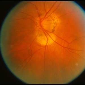
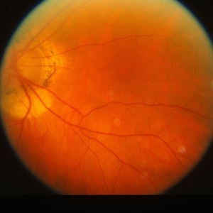
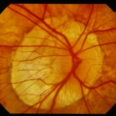
Peripapillary atrophy and central macular degeneration seen in high myopia.
Condition/keywords: high myopia, peripapillary atrophy
-
 Peripapillary Atrophy With High Myopia
Feb 4 2015 by H. Michael Lambert, MD
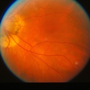
Peripapillary atrophy and central macular degeneration seen in high myopia.
Condition/keywords: high myopia, peripapillary atrophy
-
 Perpapillary Atrophy
Jul 31 2013 by From the Collections of Thomas M. Aaberg, MD and Thomas M. Aaberg Jr., MD
Perpapillary atrophy.
Condition/keywords: peripapillary atrophy
-
---thumb.jpg/image-square;max$300,300.ImageHandler) Perpypillary Atrophy With FA
Perpypillary Atrophy With FA
Jul 31 2013 by From the Collections of Thomas M. Aaberg, MD and Thomas M. Aaberg Jr., MD
Perpypillary atrophy with FA.
Condition/keywords: peripapillary atrophy
-
 Subluxation of the Lens
Dec 12 2024 by Kimberly Wakester
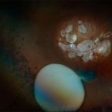
Ultra-wide field fundus photos of an 53-year-old man with a Subluxation of the Lens in the posterior vitreous cavity of the right eye after a trauma that happened many years ago. Patient remains stable with no adverse reaction to the lens at this time. No surgical intervention is recommended at this time. Patient also has myopic degeneration and lattice degeneration that will require patient to have follow up care.
Photographer: Kimberly Wakester, COA
Imaging device: Optos California
Condition/keywords: lattice degeneration, myopic degeneration, peripapillary atrophy, posterior staphyloma, Subluxation of the Lens
-
 Subluxation of the Lens
Dec 12 2024 by Kimberly Wakester
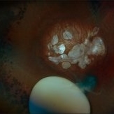
Ultra-wide field fundus photos of an 53-year-old man with a Subluxation of the Lens in the posterior vitreous cavity of the right eye after a trauma that happened many years ago. Patient remains stable with no adverse reaction to the lens at this time. No surgical intervention is recommended at this time. Patient also has myopic degeneration and lattice degeneration that will require patient to have follow up care.
Photographer: Kimberly Wakester, COA
Imaging device: Optos California
Condition/keywords: lattice degeneration, myopic degeneration, peripapillary atrophy, posterior staphyloma, Subluxation of the Lens
 Loading…