Search results (69 results)
-
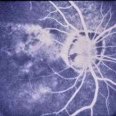
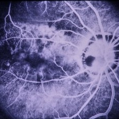
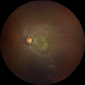
 Coloboma
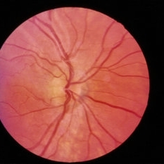
Sep 7 2018 by John S. King, MD
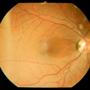
11-year-old white female with bilateral optic nerve and retinochoroidal colobomas and an optic nerve pit in the right eye looking almost like pseudoduplication of the optic nerve. She is currently 20/30 OD and 20/20 OS. She has a history of laser by Dr. Zocchi about 10 years ago for a low lying, macula involving, serous retinal detachment, and has responded well.
Photographer: Stacey Coleman
Imaging device: Topcon
Condition/keywords: chorioretinal coloboma, inferior optic nerve coloboma, optic disc pit
-
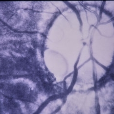
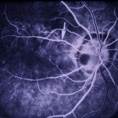
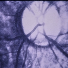
 Color Fundus Photograph of Right Optic Disc Pit
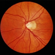
Jul 20 2019 by Arwa Azmeh, MD, PhD
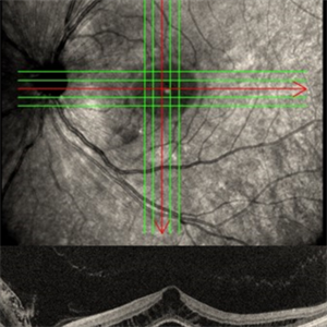
Fundus photograph of 38-year-old healthy man with right optic disc pit, who recently noticed slightly blurred vision in right eye while closing the left eye. BCVA was 20/25 in OD and 20/20 in OS. IOP was 15mmHg OD and 14 mmHg OS. Right fundus exam showed small optic disc pit near the temporal rim of optic disc with abnormal reflex of nasal macula. Left fundus was normal. Late FA of right optic disc showed no leakage or staining of optic disc. Macular OCT showed normal foveal contour with no subretinal fluid or macular edema. There was significant reduction in RNFL thickness in the temporal sector in right eye. Coloboma is clearly seen on vertical OCT scan as well as horizontal scans through right optic pit.
Photographer: Ebtisam Aljbeili, Damascus university, Almouassat university hospital
Imaging device: Heidelberg Spectralis 2
Condition/keywords: optic disc pit
-
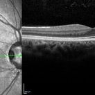
 Left Eye Optical Coherence Tomography Showing Optic Disc Pit
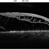
Nov 9 2024 by Anand Temkar
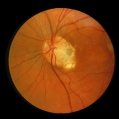
Left Eye Optical Coherence Tomography of a 48 years old male patient showing Optic Disc Pit.
Photographer: Dr.Anand Temkar- Retina Foundation, Ahmedabad
Imaging device: Mirante
Condition/keywords: optic disc pit, Optic pit
-
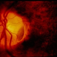
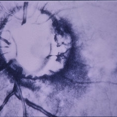
 Optic Disc Pit and CSR
Jun 4 2014 by Henry J. Kaplan, MD
Late phase angiogram clearly shows the abnormally large optic nerve with temporal pit which is stained. #4
Condition/keywords: central serous retinopathy (CSR), optic disc pit
-
 Optic Disc Pit
Jun 4 2014 by Henry J. Kaplan, MD
Optic disc pit in the temporal part of optic nerve with associated CSR.
Condition/keywords: central serous retinopathy (CSR), optic disc pit
-
 Optic Disc Pit
Dec 11 2014 by H. Michael Lambert, MD
Fluorescein angiogram of RE with ON pit and macular detachment with RPE mottling.
Condition/keywords: optic disc pit
-
 Optic Disc Pit
Dec 11 2014 by H. Michael Lambert, MD
High mag red free of RE optic nerve pit.
Condition/keywords: optic disc pit
-
 Optic Disc Pit
Dec 11 2014 by H. Michael Lambert, MD
High mag color photograph of LE optic nerve pit.
Condition/keywords: optic disc pit
-
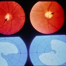
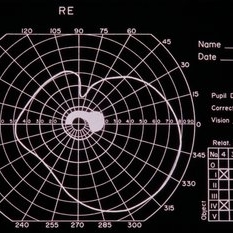 Optic Disc Pit
Dec 11 2014 by H. Michael Lambert, MD
Goldmann VF of RE centrocecal VF defect due to ON pit.
Condition/keywords: optic disc pit
-
 Optic Disc Pit
Dec 11 2014 by H. Michael Lambert, MD
High mag red free of LE optic nerve pit.
Condition/keywords: optic disc pit
-
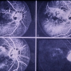
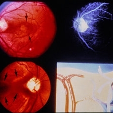 Optic Disc Pit
Dec 11 2014 by H. Michael Lambert, MD
4 image montage of ON pit with RE and LE color disc photo, LE angiogram and schematic artist drawing.
Condition/keywords: optic disc pit
-
 Optic Disc Pit
Dec 11 2014 by H. Michael Lambert, MD
Optic Disc Pit, color photo RE.
Condition/keywords: optic disc pit
-
 Optic Disc Pit
Dec 11 2014 by H. Michael Lambert, MD
Early phase FA leakage from ON pit RE.
Condition/keywords: optic disc pit
-
 Optic Disc Pit
Dec 11 2014 by H. Michael Lambert, MD
Late phase FA of leakage from ON pit RE.
Condition/keywords: optic disc pit
-
 Optic Disc Pit
Dec 11 2014 by H. Michael Lambert, MD
4 image montage of ON pit with RE and LE color disc photo, RE and LE.
Condition/keywords: optic disc pit
-
 Optic Disc Pit
Dec 11 2014 by H. Michael Lambert, MD
High mag red free of LE optic nerve pit.
Condition/keywords: optic disc pit
-
 Optic Disc Pit
Dec 11 2014 by H. Michael Lambert, MD
4 image montage of early to late phase FA showing leakage from ON pit.
Condition/keywords: optic disc pit
-
 Optic Disc Pit
Dec 11 2014 by H. Michael Lambert, MD
High mag RE color photo of ON pit.
Condition/keywords: optic disc pit
-
 Optic Disc Pit
Dec 11 2014 by H. Michael Lambert, MD
High mag RE color photo of ON pit.
Condition/keywords: optic disc pit
-
 Optic Disc Pit
Dec 11 2014 by H. Michael Lambert, MD
High mag LE color photo of ON pit.
Condition/keywords: optic disc pit
-
 Optic Disc Pit
Dec 11 2014 by H. Michael Lambert, MD
High mag LE color photo of ON pit.
Condition/keywords: optic disc pit
-
 Optic Disc Pit
Oct 8 2019 by DIEGO TOLENTINO
OCT of a patient with optic disc pit.
Photographer: Diego Tolentino
Condition/keywords: optic disc pit, optical coherence tomography (OCT)
-
 Optic disc pit
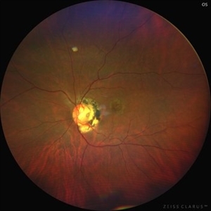
Mar 21 2022 by T. P . VIGNESH, MBBS,MS
Fundus photo of Left eye of a 55 year male patient revealing optic disc pit with temporal barrage laser marks and foveal schisis with RPE atrophic changes.
Photographer: Bharathi Singaravel
Imaging device: Zeiss Clarus
Condition/keywords: Optic disc pit
-
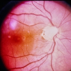
 Optic disc pit
Apr 14 2023 by T. P . VIGNESH, MBBS,MS
Fundus photograph of an 32-year-old woman with optic disc pit and macular RPE atrophy .
Photographer: Bharathi S
Imaging device: ZEISS CLARUS
Condition/keywords: Optic disc pit
-
 Optic Disc Pit
May 17 2024 by T. P . VIGNESH, MBBS,MS
SD-OCT of the right eye of a 26 year man revealing Optic disc pit .
Photographer: Sivanath
Imaging device: Heidelberg Spectralis
Condition/keywords: Optic disc pit
 Loading…