Search results (625 results)
-
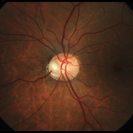
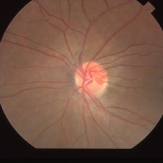 Capillary Hemangioma
Mar 27 2019 by Gary R. Cook, MD, FACS
28-year-old asymptomatic white female with a retinal capillary hemangioma of the optic disc OS; V.A.= 20/20-1.
Imaging device: Topcon VT-50
Condition/keywords: optic disc
-
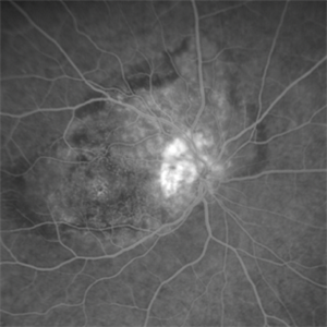
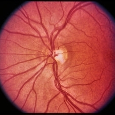
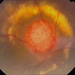 Color Photo of Optic Disc Capillary Hemangioblastoma
Mar 18 2014 by Arwa Azmeh, MD, PhD
Color fundus photograph of an 48-year-old male who complained of decreased visual acuity in his right eye over the last few months. Systemically the patient was healthy. His VA was OD Cf 3m, OS 20/20. Anterior segments were WNL in OU. IOP was WNL in OU. Fundus exam OD revealed unpigmented mass over the optic disc with retinal venous tortuosity at its edges with a ring of thick HYE surrounding it and shallow RD in this area extending to the foveal area. Several few small retinal hemorrhages were seen in the far retinal periphery which were explained to be caused by venous stasis due the optic disc tumor.
Condition/keywords: color photo, optic disc, retinal hemangioblastoma
-
 Congenital Hypertrophy of the Retinal Pigment Epithelium
Nov 11 2019 by Jessica Norkus
Bilateral Optos ultra wide field imaging of a 31-year-old female patient with CHRPE lesions. Lesions in OD were suspicious of Gardner Syndrome due to familial history of cancerous polyps in colon. Patient underwent colonoscopy and was deemed clear.
Photographer: Jessica Norkus, COA, Retina Specialists of Michigan
Imaging device: Optos Ultra Wide Field Camera
Condition/keywords: bear tracks, bilateral, color fundus photograph, color photo, congenital hypertrophy of the retinal pigment epithelium (CHRPE), fundus autofluorescence (FAF), fundus photograph, lacunae, macula, optic disc, Optos, pseudocolor, ultra-wide field imaging
-
 Fundus Photo
Jun 6 2023 by Mayor Vang
In honor of pride month! A fundus photo using a Ziess fundus camera showing an IOL artifact.
Photographer: Mayor Vang, (OCT-C, COT), UTSW, Dallas, TX
Condition/keywords: artifact, fundus photograph, IOL, optic disc
-
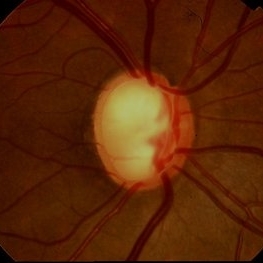
 Giant Optic Disc Melanocytoma
Jun 12 2017 by Nabil Bouslous
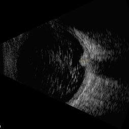
Fundus photograph, angiography and B scan of an 50-year-old woman with a giant optic disc melanocytoma which has been involving over a past year.
Photographer: Nabil Bouslous, Mohamed VI university hospital, Marrakech, Morocco.
Condition/keywords: melanocytoma, optic disc
-
 Giant Optic Disc Melanocytoma
Jun 12 2017 by Nabil Bouslous
Fundus photograph, angiography and B scan of an 50-year-old woman with a giant optic disc melanocytoma which has been involving over a past year.
Photographer: Nabil Bouslous, Mohamed VI university hospital, Marrakech, Morocco.
Condition/keywords: melanocytoma, optic disc
-
 Glaucoma
Feb 9 2015 by Govindarajan Venkatesan
Glaucoma.
Photographer: Govindarajan Venkatesan
Condition/keywords: optic disc
-
 Juxtapapillary Retinal Capillary Hemangioblastoma
Oct 16 2025 by Sara Mayoral Sánchez
A hyperfluorescent lesion located superotemporal to the optic disc, consistent with a juxtapapillary retinal capillary hemangioma.
Photographer: Sara Mayoral Sánchez, H.U.Puerta del Mar, Cádiz
Condition/keywords: angiography with fluorescein, hemangioma, optic disc, retina capillary hemangioblastoma
-
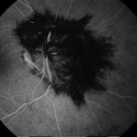
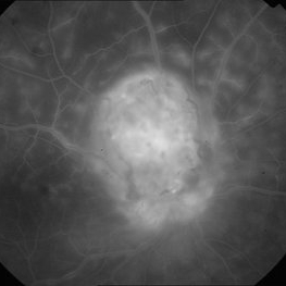 Late Phase FA of Optic Disc Capillary Hemangioblastoma
Mar 18 2014 by Arwa Azmeh, MD, PhD
Late phase FA showed increased hyper fluorescence of the mass.
Condition/keywords: optic disc, retinal hemangioblastoma
-
---thumb.jpg/image-square;max$300,300.ImageHandler) Melanocytoma
Melanocytoma
Feb 13 2013 by From the Collections of Thomas M. Aaberg, MD and Thomas M. Aaberg Jr., MD
Melanocytoma, color fundus photo, optic disc.
Condition/keywords: melanocytoma, optic disc
-
 Melanocytoma of the disc
Dec 27 2023 by NIDHI PANWAR, MD FRCS Glasgow FNB FICO
Fundus photograph of an otherwise healthy 41 year old female , with recently detected diabetes mellitus and came for fundoscopy was Detected to have left eye optic disc melanocytoma .
Photographer: Nidhi Panwar, NMC Royal hospital, Sharjah , UAE
Condition/keywords: melanocytoma, optic disc
-
 Morning Glory Anomaly
Mar 24 2022 by Elite Bor-Shavit, MD
Disc photo of a 28-years-old male with Morning Glory Anomaly of his right optic nerve observed over time.
Condition/keywords: Morning Glory Anomaly, optic disc
-
 Morning Glory Disc Anomaly
Aug 19 2017 by Mitzy E Torres Soriano, MD
A 10-year-old female patient with morning glory disc anomaly in her left eye.
Photographer: Mitzy E. Torres Soriano
Condition/keywords: coloboma of optic disc, coloboma of the optic nerve, Morning Glory Syndrome, optic disc, optic disc dysplasia
-
 Nasal Optic Disc Pit
May 3 2022 by Bernardo Araújo
Asymptomatic patient. 41-year-old woman.
Photographer: Bernardo Araújo, Retina Clinic, São Paulo, SP, Brazil.
Condition/keywords: nasal, optic disc
-
---thumb.jpg/image-square;max$300,300.ImageHandler) Optic And Hyperemic Disc And Hyperemic Rim
Optic And Hyperemic Disc And Hyperemic Rim
Nov 4 2013 by Maurice F. Rabb
Color photographs of the right posterior pole demonstrates an optic disc with a cup/disc ratio of 0.5 and a hyperemic disc and hyperemic rim of neutral tissue.
Condition/keywords: hyperemic disc, hyperemic rim, optic disc
-
 Optic Disc
Dec 18 2014 by H. Michael Lambert, MD
Image of optic disc OS in a 15-year-old white female with peau d'orange fundus changes but biopsy negative for PXE.
Condition/keywords: optic disc, peau d'orange fundus
 Loading…