Search results (70 results)
-
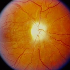
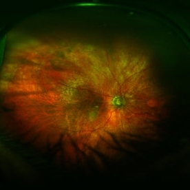
 BRVO With Optic Atrophy
Nov 27 2020 by Sham Talati, DOMS
A 55-year-old male patient presented with glaucomatous optic atrophy with ST BRVO in the right eye.
Photographer: Dr. Sham Talati,Retina Foundation,Ahmedabad
Imaging device: Nidek Mirante
Condition/keywords: branch retinal vein occlusion (BRVO), branch vein occlusion (BVO), glaucomatous atrophy of optic disc, optic atrophy
-
 Disseminated Chorioretinitis With Unknown Etiology
Apr 5 2018 by Kim Barrett
Ultra-wide field fluorescein angiogram of a 31-year-old female with intermittent pain in her left eye. Her condition has been managed in Liberia until recently when she moved to the United States. She suffers from multiple modalities including central retinal artery occlusion, posterior synechiae of the iris, interstitial keratitis, disseminated chorioretinitis, as well as HIV. An infectious cause is high on the differential in light of her HIV status. DDx: hypertensive crisis, an embolism (? IV drug use), coagulopathy, trauma, infectious. Blood work was normal. Her current vision is 20/30 right eye and 20/400 left eye.
Photographer: Kim Barrett, COA
Imaging device: Optos
Condition/keywords: central retinal artery occlusion (CRAO), chorioretinal scar, ciliary artery sparring, disseminated chorioretinitis, HIV, left eye, optic atrophy, staining
-
 Extensive/ Heavy Focal/ PRP OD - Color
Jun 28 2018 by Hosam Attia, MD
70-year-old woman, seen for initial eye exam, with endstage PDR and H/O prior Focal/ PRP OU , somewhere else.
Imaging device: Optos - California
Condition/keywords: chorioretinal scar, focal laser, ghost vessels, optic atrophy, pan-retinal photocoagulation (PRP), proliferative diabetic retinopathy (PDR)
-
 Extensive/ Heavy Focal/ PRP OD - FAF
Jun 28 2018 by Hosam Attia, MD
70-year-old woman, seen for initial eye exam, with endstage PDR and H/O prior Focal/ PRP OU , somewhere else.
Imaging device: Optos - California
Condition/keywords: chorioretinal scar, focal laser, ghost vessels, optic atrophy, pan-retinal photocoagulation (PRP), proliferative diabetic retinopathy (PDR)
-
 Extensive/ Heavy Focal/ PRP OS - Color
Jun 28 2018 by Hosam Attia, MD
70-year-old woman, seen for initial eye exam, with endstage PDR and H/O prior Focal/ PRP OU , somewhere else.
Imaging device: Optos - California
Condition/keywords: chorioretinal scar, focal laser, ghost vessels, optic atrophy, pan-retinal photocoagulation (PRP), proliferative diabetic retinopathy (PDR)
-
 Extensive/ Heavy Focal/ PRP OS - FAF
Jun 28 2018 by Hosam Attia, MD
70-year-old woman, seen for initial eye exam, with endstage PDR and H/O prior Focal/ PRP OU , somewhere else.
Imaging device: Optos - California
Condition/keywords: chorioretinal scar, ghost vessels, laser scarring, optic atrophy, pan-retinal photocoagulation (PRP), proliferative diabetic retinopathy (PDR), retinal scar
-
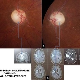 Glioblastoma Multiform Causing Bilateral Optic Atrophy
Mar 8 2021 by Deepak Bhojwani, MS
A 27-year-old male presented with complaints of severe headache associated with progressive loss of vision in both eyes since last 2- 3 months. On ocular examination his visual acuity was no perception of light in both eyes with bilateral dilated pupils not reacting to light. Fundus examination revealed bilateral optic atrophy (Figures A &B). He was advised an MRI scan for neurological evaluation. The patient was advised to review with Neurologist after getting MRI scans and review again with us. To our surprise this young patient was harboring a massive brain tumor reported as glioblastoma multiforme by the radiologist (Figure C). He has been advised tumor resection and chemoreduction by the neuro-oncologist. This case highlights neurological evaluation of all patients with bilateral optic atrophy. The presenting complaint of headache also prompted us for getting neurological examination. So its rightly said : eyes are the window to the body.
Photographer: DEEPAK BHOJWANI
Imaging device: ZEISS VISUCAM 524
Condition/keywords: optic atrophy
-
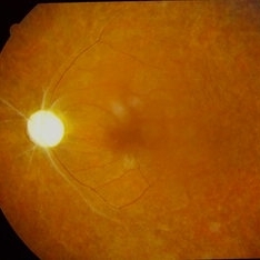 Optic atrophy
Sep 14 2023 by Ben Serar
Fundus photograph of LE showing Pale disc in a case of optic atrophy, with arteriolar attenuation with vascular sheathing and granular fundus.
Condition/keywords: optic atrophy
-
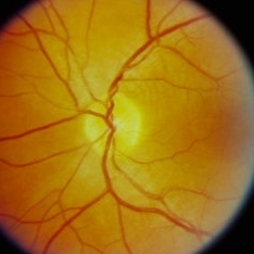 Optic Atrophy
Sep 14 2023 by Ben Serar
Fundus photograph of the LE showing disc pallor with well-defined disc margins in a case of optic atrophy.
Condition/keywords: optic atrophy
-
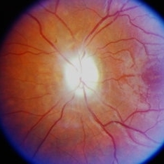 Optic Atrophy
Sep 12 2023 by Ben Serar
Fundus photograph of the LE showing disc pallor with hazy disc margins in a case of secondary optic atrophy
Condition/keywords: optic atrophy
-
 Optic Atrophy
Sep 12 2023 by Ben Serar
Fundus photograph of the RE showing disc pallor with hazy disc margins in a case of secondary optic atrophy
Condition/keywords: optic atrophy
-
 Optic Atrophy
Sep 12 2023 by Ben Serar
Fundus photograph of the LE showing disc pallor with well-defined disc margins in a case of primary optic atrophy
Condition/keywords: optic atrophy
-
 Optic atrophy
Apr 23 2015 by Mehul A Shah
23-year-old male presented with sudden loss of vision following methyl alcohol poisoning and picture after 3 months.
Photographer: Mehul Shah
Imaging device: Zeiss FF450 Plus
Condition/keywords: optic atrophy
-
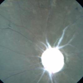
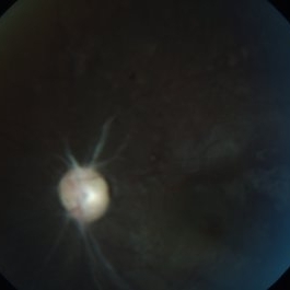
 Optic Atrophy
May 2 2013 by Henry J. Kaplan, MD
Left optic atrophy as a chalky white optic nerve.
Condition/keywords: optic atrophy
-
 Optic Atrophy and Attenuated Retinal Vessels Following Endophthalmitis
Jul 12 2014 by Philip J. Polkinghorne, MD
This elderly lady underwent a vitrectomy for post-surgical endophthalmitis. The infection was successfully treated but the functional outcome was poor because of optic atrophy and attenuated retinal vessels.
Photographer: Alex Fraser
Imaging device: Optos Camera
Condition/keywords: attenuated vessels, endophthalmitis, optic atrophy, post-vitrectomy
-
 Optic Atrophy With Pigmented Epithellium
Apr 16 2014 by Dipankar Barua, M.Sc
Female patient, 22-years-old. On examination her vision in the right eye is no perception of light and left eye is 6/6. The right upper has orbital growth. It seems to be a case of Optic atrophy with pigmented epithellium.
Photographer: Dipankar Barua
Imaging device: Topcon TRC 50 DX (IA)
Condition/keywords: optic atrophy, pigment epithelial detachment (PED)
-
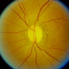
 Optic atropy
Feb 9 2015 by Govindarajan Venkatesan
Optic atrophy.
Photographer: Govindarajan Venkatesan
Condition/keywords: optic atrophy
-
 Pale Optic Atrophy
Apr 16 2014 by Dipankar Barua, M.Sc
Female patient, 16-years-old. On examination her vision of right eye has no perception of light & left eye has counting finger 1t 3 meter. She has history of surgery for craniopharyngioma. It seems to be a case of pale optic atrophy.
Photographer: Dipankar Barua
Imaging device: Topcon TRC 50 DX (IA)
Condition/keywords: optic atrophy
-
 Pale Optic Atrophy
Apr 16 2014 by Dipankar Barua, M.Sc
Female patient, 16-years-old. On examination her vision of right eye has no perception of light and left eye has counting finger 1t 3 meter. She has history of surgery for craniopharyngioma. It seems to be a case of pale optic atrophy.
Photographer: Dipankar Barua
Imaging device: Topcon TRC 50 DX (IA)
Condition/keywords: optic atrophy
-
 Post Papillitic Optic Atrophy
Sep 10 2014 by Mehul A Shah
35-year-female patient presented with progressive loss of vision following.
Photographer: Drashti Netralaya
Imaging device: Zeiss ff450
Condition/keywords: optic atrophy
-
 Pseudo Foster Kennedy Syndrome
Oct 13 2022 by Aditya S Kelkar, MS, FRCS, FASRS,FRCOphth
Colour fundus photograph of a 44-year-old man showing bilateral small discs with optic atrophy on the right eye and disc edema on the left eye resulting from consecutive NAAION in both eyes.
Photographer: Dr Sukanya Mondal, National Institute of Ophthalmology, Pune. India
Imaging device: Zeiss Clarus 500
Condition/keywords: ischemic optic neuropathy, optic atrophy, optic disc edema
-
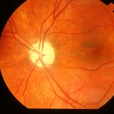
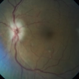
 Secondary Optic Atrophy
Sep 20 2014 by Mehul A Shah
A 24-year-old male presented with complaint of loss of vision, on examination he was found to have this picture.
Photographer: Drashti Netralaya,Dahod
Imaging device: Zeiss ff450
Condition/keywords: optic atrophy
-
 Secondary Optic Atrophy
Oct 2 2017 by Mehul A Shah
A patient presented after operated for pitutary adenoma and complained of loss of vision this is clinical picture.
Photographer: Mehul Shah
Condition/keywords: optic atrophy
-
 Slide 11-27
Feb 26 2019 by Lancaster Course in Ophthalmology
Optic atrophy. A normal optic nerve ( x 16). (Scheie Eye Institute, No. 2333.)
Condition/keywords: optic atrophy, optic nerve
-
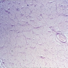
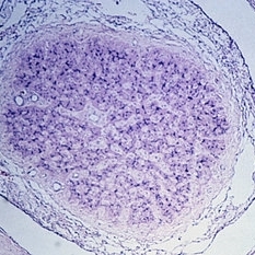 Slide 11-28
Feb 26 2019 by Lancaster Course in Ophthalmology
Optic atrophy taken at the same magnification to show marked shrink age. Note that the optic sheath appears to be enlarged. The nerve is almost completely replaced by connective tissue. (Scheie Eye Institute, No. 3917.)
Condition/keywords: optic atrophy
 Loading…