Search results (9 results)
-
 Atypical Tubercular Occlusive Peripheral Retinal Vasculitis
Jun 21 2024 by Tejaswita Verma
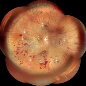
Follow up right eye fundus photograph of a 27 year old male with vision 6/12 , diagnosed with systemic tuberculosis(mediastinal lymphadenopathy on chest CT) on oral steroids, and started on ATT .We can see a parafoveal sub-ILM hemorrhage with vascular sheathing and hemorrhages in inferior and temporal quadrants . The patient was advised anti-VEGF intravitreal injection, later sectoral laser after resolution of inflammation
Photographer: DR. TEJASWITA VERMA
Imaging device: MIRANTE
Condition/keywords: obliterative peripheral vasculitis, ocular tuberculosis
-
 Atypical Tubercular Peripheral Occlusive Retinal Vasculitis
Jun 21 2024 by Tejaswita Verma
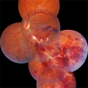
Fundus montage of the right eye of a 27 year old male with macula threatening occlusive vasculitis showing hemorrhages in inferior, temporal quadrant with vascular sheathing. The patient was Mantoux positive (20 mm induration) and IGRA (TB-GOLD)positive and started on oral steroids. The case was atypical due to no vitritis at presentation which is unusual of tuberculosis. Behcet's disease was ruled out as there was no panuveitis like picture.
Photographer: DR. TEJASWITA VERMA
Imaging device: MIRANTE
Condition/keywords: occlusive vasculitis, ocular tuberculosis
-
 Choroidal Tubeculoma
Feb 12 2021 by Sham Talati, DOMS
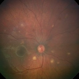
A 9-year-old male patient who is a known case of pulmonary tuberculosis presented with choroidal tubeculoma in his right eye.
Photographer: Dr. Sham Talati,Retina Foundation,Ahmedabad
Imaging device: Nidek Mirante
Condition/keywords: choroidal tuberculoma, ocular tuberculosis, tuberculosis
-
 Choroidal Tuberculoma Fluorescence Angiography Montage
Feb 12 2021 by Sham Talati, DOMS
A 9-year-old male patient who is a known case of pulmonary tuberculosis presented with choroidal tuberculoma in his right eye.
Photographer: Dr. Sham Talati,Retina Foundation,Ahmedabad
Imaging device: Nidek Mirante
Condition/keywords: choroidal tuberculoma, ocular tuberculosis, tuberculosis
-
 FFA in Atypical Tubercular Peripheral Occlusive Retinal Vasculitis
Jun 21 2024 by Tejaswita Verma
Right eye FFA montage of a 27 year male with peripheral occlusive tubercular vasculitis, showing CNP areas inferiorly and temporally, leakages and blocked fluorescence due to hemorrhages. The patient was advised intravitreal anti-VEGF injection and later sectoral laser once inflammation subsides.
Photographer: DR. TEJASWITA VERMA
Imaging device: MIRANTE
Condition/keywords: obliterative peripheral vasculitis, ocular tuberculosis
-
 Miliary Tuberculosis
Mar 17 2022 by Franco Benvenuto, MD
Fundus photograph of a 9-month-old baby with hemophagocytic syndrome secondary to Tuberculosis infection.
Photographer: Franco Benvenuto, Universidad de Buenos Aires, Argentina
Imaging device: RetCam
Condition/keywords: ocular tuberculosis
-
 Ocular Tuberculosis
May 18 2020 by McGill University Health Centre
Ocular tuberculosis refers to necrotizing granulomatous uveitis caused by Mycobacterium tuberculosis infection. The mechanism of infection of ocular structures is via hematogenous dissemination of the bacteria during the primary infection. In this enucleation specimen, the anterior chamber and vitreous cavity are filled by caseous exudate. The iris is also replaced by a whitish material, and the retina is completely detached. Note the whitish thickening of the posterior choroid (arrow).
Condition/keywords: caseous exudate, enucleation, ocular tuberculosis, uveitis
-
 Serpiginous-Choroiditis-Like
Mar 30 2024 by Karen Flores Guevara
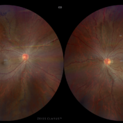
Fundus photograph of a 26-year-old woman with a serpiginous-choroiditis-like presentation secundary to a tuberculosis activation disease, started with visual acuity and systemic syntomps
Photographer: Andrés Santiago Pérez-Giraldez, MD. Asociación para evitar la Ceguera en Mexico I.A.P. Mexico
Condition/keywords: ocular tuberculosis, serpiginous choroiditis, tuberculosis
-
 Tubercular Retinal Vasculitis
Mar 31 2022 by Lucas Zago Ribeiro, MD
Fundus image of 22yr male, presenting with unilateral vision loss
Photographer: Lucas Zago Ribeiro, UNIFESP / EPM
Imaging device: Zeiss Visucam 524
Condition/keywords: ocular tuberculosis, tuberculosis, vasculitis
 Loading…