Search results (8 results)
-
 Central Retinal Vein Occlusion
Jan 21 2022 by Olivia Rainey
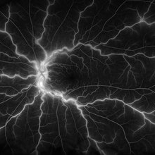
Ultra-widefield fluorescein angiogram of a 23-year-old female with a Central Retinal Vein Occlusion affecting her left eye. The patient presented on 12/22/2021 cc20/40-2 vision in the left eye. The patient reported recent trauma of being hit with a fist on both sides of face followed by vision loss. The patient has history of Hashimoto's thyroid disease. The following labs have been ordered, PT, PTT, CBC, antithrombin III activity, protein C, protein S, Factor V Leiden mutation, Prothrombin (G20210A), lipid panel, HbA1c, quantiferon gold, RPR, and CXR.
Photographer: Olivia Rainey, OCT-C, COA
Imaging device: Optos California
Condition/keywords: central retinal vein occlusion (CRVO), disc leakage, fluorescein angiogram (FA), fluorescein leakage, left eye, non-ischemic central retinal vein occlusion (CRVO), Optos, trauma, ultra-wide field imaging
-
 CRVO Nonischemic
Jul 14 2014 by Susanna S. Park, MD, PhD
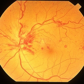
Fundus photograph of a 52-year-old woman with blurred vision showing retinal hemorrhages and some retinal venous congestion from non-ischemia central retinal vein occlusion.
Photographer: Ellen Redenbo
Condition/keywords: non-ischemic central retinal vein occlusion (CRVO)
-
 CRVO Nonischemic
Jul 9 2014 by Susanna S. Park, MD, PhD
Fundus photograph of a 60-year-old woman with new vision loss showing scattered retinal hemorrhages and retinal venous congestion with macular edema from central retinal vein occlusion.
Photographer: Ellen Redenbo
Condition/keywords: central retinal vein occlusion (CRVO), non-ischemic central retinal vein occlusion (CRVO)
-
 CRVO Nonischemic FA Vascular Staining
Jul 14 2014 by Susanna S. Park, MD, PhD
Venous transit fluorescein angiogram image of a 52-year-old woman with non-ischemia central retinal vein occlusion showing some retinal venous staining and leakage.
Photographer: Ellen Redenbo
Condition/keywords: non-ischemic central retinal vein occlusion (CRVO)
-
 CRVO with Secondary CLRAO
May 28 2020 by Richard M Martindale, MD
Non-ischemic CRVO (VA 20/30) with secondary CLRAO (nasal macular pallor) in a hypertensive 69yo female. Pathophysiologically, the cilioretinal artery occlusion occurs secondary to elevation in the hydrostatic pressure in the retinal venous system relative to the choroidal perfusion pressure (which supplies the cilioretinal artery).
Photographer: Retina Consultants of Alabama, P. C.
Imaging device: Optos
Condition/keywords: cilioretinal artery occlusion, non-ischemic central retinal vein occlusion (CRVO)
-
 Non Ischemic Hemi-CRVO
Mar 29 2013 by Henry J. Kaplan, MD
Non-ischemic CRVO: blurred disc margins, dilated and tortous veins and scattered hemorrhages in the superior half of the retina.
Condition/keywords: branch retinal vein occlusion (BRVO), central retinal vein occlusion (CRVO), hemi CRVO, non-ischemic central retinal vein occlusion (CRVO)
-
 Non-Ischemic Central Retinal Vein Occlusion
Jun 28 2016 by Nichole Lewis
53-year-old female with non-ischemic central retinal vein occlusion left eye.
Photographer: Nichole Lewis
Condition/keywords: non-ischemic central retinal vein occlusion (CRVO)
-
 Right Central Retinal Vein Occlusion
Jun 25 2021 by Ahmed Almuhaylib, MD
Fundus photograph of a 65-year-old man with uncontrolled hypertension.
Photographer: Ahmed Almuhaylib, MD, Qassim University, Kingdom of Saudi Arabia
Condition/keywords: central retinal vein occlusion (CRVO), central vein occlusion, non-ischemic central retinal vein occlusion (CRVO)
 Loading…