Search results (18 results)
-
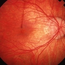
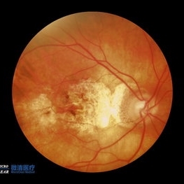
 Bilateral CNV in High Myopia
Apr 2 2019 by Gary R. Cook, MD, FACS
Right eye of a 60-year-old white female with -9D myopia, myopic maculopathy, and visible (Type 1) CNV; V.A. = 20/40.
Imaging device: Topcon VT-50
Condition/keywords: choroidal neovascular membrane (CNVM), choroidal neovascularization (CNV), high myopia, myopic degeneration, myopic fundus, pathologic myopia
-
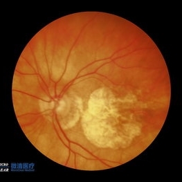
 Bilateral CNV in High Myopia
Apr 2 2019 by Gary R. Cook, MD, FACS
Left eye of a 60-year-old white female with -9D myopia and bilateral visible (Type 1) CNV; V.A. = 20/30.
Imaging device: Topcon VT-50
Condition/keywords: choroidal neovascular membrane (CNVM), choroidal neovascularization (CNV), high myopia, myopic degeneration, myopic fundus, pathologic myopia
-
 High Myopia
Apr 1 2019 by Gary R. Cook, MD, FACS
29-year-old white female with -10D high myopia OD; V.A.= 20/20.
Imaging device: Topcon VT-50
Condition/keywords: high myopia, myopic degeneration, myopic fundus
-
 High Myopia
Apr 2 2019 by Gary R. Cook, MD, FACS
51-year-old white female with -6.25D myopia OD with a myopic conus on the inferotemporal aspect of the optic disc and focal myopic chorioretinal atrophy in the macula OD; V.A. = 20/25
Imaging device: Topcon VT-50
Condition/keywords: high myopia, myopic degeneration, myopic fundus, pathologic myopia
-
 High Myopia
Apr 2 2019 by Gary R. Cook, MD, FACS
51-year-old white female with -7.00D myopia with a myopic conus on temporal aspect of the optic nerve and focal choroiretinal atrophy in the macula OS; V.A. = 20/25-1
Imaging device: Topcon VT-50
Condition/keywords: high myopia, myopic degeneration, myopic fundus, pathologic myopia
-
---thumb.jpg/image-square;max$300,300.ImageHandler) Myopic fundus
Myopic fundus
Jan 11 2013 by Hyung-Woo Kwak, MD
Myopic fundus reveals yellow-colored lacquer cracks and peripapillary atrophy. There was visible choroidal vessel due to thin retina.
Photographer: Misook Lee, Kyung Hee Univsersity Hospital, Seoul
Imaging device: Zeiss f 450 plus
Condition/keywords: myopic fundus
-
 Myopic macular degeneration
Jan 11 2013 by Alex P. Hunyor, MD
Myopic macular degeneration, left eye - extensive chorioretinal atrophy.
Condition/keywords: myopic degeneration, myopic fundus, myopic macular degeneration
-
 Sneaky bubble under the buckle
May 19 2022 by Priyanka Raj, MBBS, MS
First post-operative day picture of a highly myopic eye of a patient who underwent superior 120 degrees 276 circumferential scleral buckle for bullous retinal detachment. The picture shows a remnant air bubble used as a tamponade, restricted by the buckle height and a break treated with cryopexy, well supported by the buckle.
Photographer: Ajeet, Prakash Netra Kendra
Imaging device: Zeiss Clarus 500
Condition/keywords: Cryopexy, high myopia, myopic fundus, retinal tear, scleral buckle
-
 Myopic Fundus With Staphyloma and a Peripheral Lasered Hole
Jul 25 2019 by Manish Nagpal, MD, FRCS (UK), FASRS
Wide field view of a myopic fundus with peripheral lasered hole with pigmentation along with staphyloma in the central area.
Photographer: Gayathri Mohan
Condition/keywords: laser photocoagulation, myopia, ultra-wide field imaging
-
 CSNB-OCT-OD
Aug 23 2021 by Jennifer Carstens
OCT/infrared image showing myopic fundus with normal retinal structure in patient with CACNA1F associated X-linked CSNB (OD).
Photographer: Jing Zhang, Ophthalmic Photographer
Condition/keywords: congenital stationary night blindness (CSNB), infrared image, optical coherence tomography (OCT)
-
 CSNB-OCT-OS
Aug 23 2021 by Jennifer Carstens
OCT/infrared image showing myopic fundus with normal retinal structure in patient with CACNA1F associated X-linked CSNB (OS).
Photographer: Jing Zhang, Ophthalmic Photographer
Condition/keywords: congenital stationary night blindness (CSNB), infrared image, optical coherence tomography (OCT)
-
 Fundus Albipunctata
Dec 27 2016 by Elad Moisseiev, MD
A 53-year-old female patient with high myopia and complaints of stationary night blindness since childhood. Fundus: myopic fundus with yellow dots in the posterior pole. Genetics: Homozygous mutations in RDH5 gene - c.160C>T (p.R54X), confirming the diagnosis of fundus albipunctata.
Photographer: Galit Yair-Pur
Condition/keywords: fundus albipunctatus
-
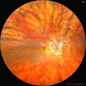 Myopic CNVM
Jul 22 2022 by T. P . VIGNESH, MBBS,MS
Fundus photograph of a 64-year-old woman with high myopia , myopic fundus and myopic CNVM.
Photographer: Bharathi Singaravel
Imaging device: Zeiss Clarus
Condition/keywords: high myopia, Myopic CNVM
-
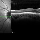
 Myopic Foveoschisis
May 19 2014 by Ahmad Gawady
OCT Of 56-year-old highly myopic female with drop of vision BCVA 2/60. Clear media, myopic fundus , Ill defined macular abnormality. OCT shows inferior juxtafoveal splitting of Inner retinal layer. No evidence of PVD, CNV or leakage .
Condition/keywords: myopic foveoschisis, optical coherence tomography (OCT)
-
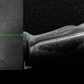
 Myopic Foveoschisis
May 19 2014 by Ahmad Gawady
OCT Lt of a 56-year-old female with drop of vision OS bilateral high myopia -6.0 D. Normal I.O.P., BCVA 6/18 OD , 2/60 OS., clear media, myopic fundus OD. No abnormalities OD, myopic fundus Ill defined fovea abnormality OS. OCT macular area OS : inferior juxtafoveal splitting of inner retinal layer (myopic foveoschisis) No evidence of PVD, CNV or leakage.
Condition/keywords: high myopia, optical coherence tomography (OCT)
-
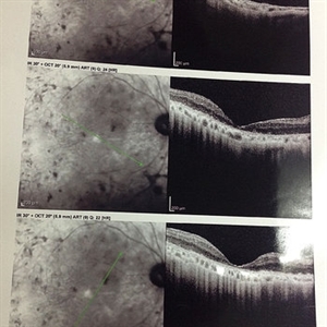
 Myopic Macular Degeneration
Oct 21 2022 by Vaibhavi Noticewala, M S Ophthalmology, FVRS
RE Myopic fundus with Myopic macular degeneration with foster fuch spot and Active CNVM with glaucomatous disc cupping
Photographer: Optom Priyanshi Kambodi
Condition/keywords: Myopia macular degeneration CNVM foster fuch spot
-
 Myopic Macular Degeneration
Oct 21 2022 by Vaibhavi Noticewala, M S Ophthalmology, FVRS
LE Myopic fundus with Myopic macular degeneration and Active CNVM with glaucomatous disc cupping
Photographer: Optom Priyanshi Kambodi
Condition/keywords: Myopia macular degeneration CNVM
-
Smartphone Fundoscopy
Apr 26 2023 by Kalyan Singh
Low cost, portable, high-tech technique of fundoscopy using a smartphone with 20 D or 2.2 panretinal auxiliary lens. This video showing a myopic fundus.
Condition/keywords: fundoscopy, myopia, smartphone
 Loading…