Search results (103 results)
-
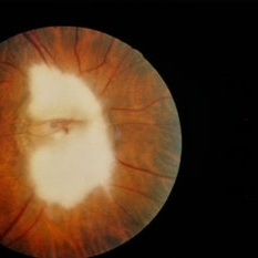
 Myelinated Nerve Fiber
Sep 21 2014 by Mehul A Shah
A 35-year-old female came for refraction and on examination myelinated nerve fibers detected.
Photographer: Drashti Netralaya,Dahod
Imaging device: Zeiss ff450
Condition/keywords: myelinated nerve fibers
-
---thumb.jpg/image-square;max$300,300.ImageHandler) Myelinated Nerve Fiber
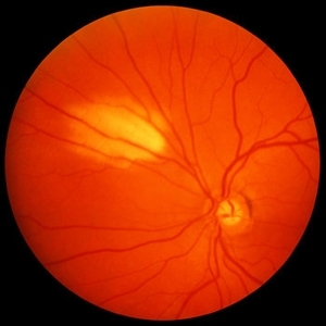
Myelinated Nerve Fiber
Feb 20 2013 by From the Collections of Thomas M. Aaberg, MD and Thomas M. Aaberg Jr., MD
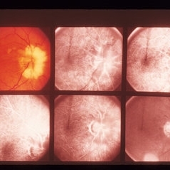
Photo of an eye with myelinated nerve fiber around the optic nerve taken with a filter to show pseudofluorescence.
Condition/keywords: myelinated nerve fibers, pseudofluorescence
-
 Myelinated Nerve Fiber
May 5 2021 by Priya Rasipuram Chandrasekaran, MBBS, DO, DNB, FRCS
A 31-year-old male presented with a decreased vision of 20/125 N24 with -6.50 DS/-3.50 cyl 90 in the left eye. Fundus examination revealed peripapillary MNF progressing superiorly, obscuring disc and vessels and sparing the macula. OCT of ONH showed hyper reflective NFL and an abrupt ending of RPE and inner retinal layers (IRL) with underlying shadowing at the beginning of hyper reflectivity. The absence of photoreceptor integrity line (PIL) in the macula is believed to cause refractory amblyopia in such patients.
Condition/keywords: myelinated nerve fibers
-
 Myelinated Nerve Fiber
Nov 10 2022 by Tandava Krishnan
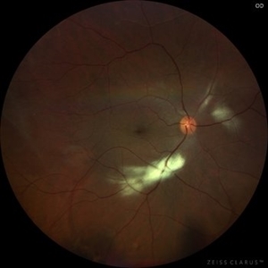
Color fundus photo of the Left eye of a patient showing myelinated nerve fiber
Condition/keywords: myelinated nerve fibers
-
 Myelinated Nerve Fibre (MNF)
Sep 12 2023 by Ben Serar
Fundus photograph of RE showing Myelinated Nerve Fibre along superior disc margin
Condition/keywords: MNF, myelinated nerve fiber
-
 AO
AO
Sep 3 2013 by Howard Schatz, MD
Myelinated nerve fibers, AO, BAO.
Condition/keywords: AO, myelinated nerve fibers
-
---thumb.jpg/image-square;max$300,300.ImageHandler) ARMD RPE Defect / Myelinated NFL
ARMD RPE Defect / Myelinated NFL
Jan 9 2014 by David Callanan, MD
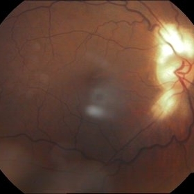
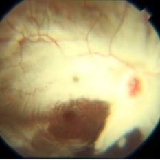
ARMD RPE defect , myelinated nerve fiber layer in a 47-year-old male patient.
Condition/keywords: myelinated nerve fiber layer, retinal pigment epithelium (RPE) defect
-
---thumb.jpg/image-square;max$300,300.ImageHandler) ARMD RPE Defect / Myelinated NFL
ARMD RPE Defect / Myelinated NFL
Jan 9 2014 by David Callanan, MD
ARMD RPE defect , myelinated nerve fiber layer in a 47-year-old male patient.
Condition/keywords: myelinated nerve fiber layer, retinal pigment epithelium (RPE) defect
-
 Isolated myelinated nerve fiber layers
Mar 5 2023 by Niloofar Piri, MD
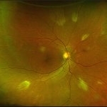
Fundus photograph of the right eye demonstrating patches of isolated myelinated nerve fiber layers along inferior arcade as well as nasal retina
Photographer: Sean Kelso, Saint Louis University
Condition/keywords: myelinated nerve fiber layer, myelinated nerve fibers
-
 Medullated nerve fibres
Mar 5 2023 by Kalyan Singh
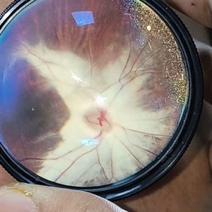
History of trauma 4-5 years back and presented to our side with unilateral diminution of vision.
Photographer: Kalyan Singh, GSVM medical college, Kanpur
Imaging device: Smartphone (1 plus 10 R)
Condition/keywords: myelinated nerve fibers, trauma
-
 MNFL
MNFL
Sep 5 2015 by Ali Tavallali, MD, FASRS
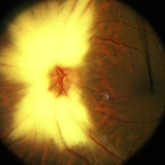
A 16-year-old male with diffuse MNFL of OD.
Photographer: Neda Shaibani
Condition/keywords: myelinated nerve fiber layer
-
 MNFL
MNFL
Sep 5 2015 by Ali Tavallali, MD, FASRS
A 16-year-old malewith diffuse MNFL of OD.
Photographer: Neda Shaibani
Condition/keywords: myelinated nerve fiber layer
-
 Multiple Areas of Myelinated RNFL OD
Sep 18 2019 by John S. King, MD
68-year-old African American male presented with an acute PVD in the fellow eye. Fellow eye had similar findings, but the pics were not as good as OD.
Photographer: Brittany Dewberry
Imaging device: Optos CA
Condition/keywords: myelinated nerve fiber layer, myelinated nerve fibers
-
 Myelianated Nerve Fiber Layer
May 2 2013 by Henry J. Kaplan, MD
Extensive myelinated nerve fibers.
Condition/keywords: myelinated nerve fibers
-
 Myelinated Nerve Fiber (mNFL)
Jun 21 2020 by Dhaivat Shah
Myelinated nerve fiber layer (mNFL) is a benign clinical entity that results from an embryologic developmental anomaly. Myelination along the visual pathway is noted around the eighth month of gestation, and typically reaches the posterior globe around the time of birth with virtually all fibers reaching complete myelination by age 7 months till the lamina cribrosa. Sometimes, due to altered neuro hormonal signals, this process of myelination extends past the lamina cribrosa and is visible on fundus examination as distinct white patches on the inner retinal surface. On infrared and red-free imaging, mNFL appears white, which is likely due to the high lipid content of myelin. Myelin blocks detection of underlying fluorescent material, thus appearing dark on fundus autofluorescence. On optical coherence tomography , it appears as a thickened and hyperreflective retinal nerve fiber layer. mNFL is typically benign but can be mistaken for other potentially serious conditions like neoplastic infiltration or infection. Hence, it is crucial to recognize the benign nature of mNFL to avoid superfluous medical testing.
Photographer: Ms Srishti Sharma
Imaging device: Choithram Netralaya
Condition/keywords: myelinated nerve fibers
-
---thumb.jpg/image-square;max$300,300.ImageHandler) Myelinated Nerve Fiber Layer
Myelinated Nerve Fiber Layer
Feb 20 2013 by From the Collections of Thomas M. Aaberg, MD and Thomas M. Aaberg Jr., MD
Myelinated nerve fiber layer Optic nerve Fundus Photo
Condition/keywords: myelinated nerve fibers
-
 Myelinated Nerve Fiber Layer
Aug 2 2020 by Kelly Hannan
Myelinated nerve fiber layer.
Imaging device: heidelberg spectralis
Condition/keywords: myelinated nerve fiber layer
-
 Myelinated Nerve Fiber Layer
Nov 24 2022 by Eder Díaz Dorado
Fundus photograph of an 35-year-old woman with myelinated nerve fiber layer
Photographer: Eder Díaz Dorado, Hospital Central Militar CDMX
Imaging device: Smartphone
Condition/keywords: myelinated nerve fibers
-
 Myelinated Nerve Fiber Layer
Jan 30 2015 by H. Michael Lambert, MD
Myelin of optic nerve head covering optic nerve grossly.
Condition/keywords: myelinated nerve fiber layer
-
 Myelinated Nerve Fiber Layer
Mar 26 2019 by Gary R. Cook, MD, FACS
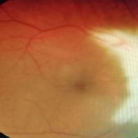
39-year-old white male with persistent myelination of the nerve fiber layer superiorly in his left eye; VA=20/20.
Imaging device: Topcon VT-50
Condition/keywords: myelinated nerve fiber layer
-
 Myelinated Nerve Fiber Layer
Oct 8 2012 by Jeffrey G. Gross, MD, FASRS
Myelinated nerve fiber layer superonasal retina.
Condition/keywords: myelinated nerve fibers, superonasal retina
 Loading…