Search results (15 results)
-
 Breast cancer metastatic to choroid
Jul 13 2021 by Odette M. Houghton, MD
A-scan image of a 59-year-old female with a choroidal tumor secondary to metastatic breast cancer.
Photographer: Christina Carpenter COA, ROUB, OSC, Mayo Clinic Arizona
Imaging device: Ellex
Condition/keywords: a-scan image, breast cancer, metastatic cancer
-
 Breast cancer metastatic to choroid
Jul 13 2021 by Odette M. Houghton, MD
B-scan image of a 59-year-old female with a choroidal tumor secondary to metastatic breast cancer.
Photographer: Christina Carpenter COA, ROUB, OSC, Mayo Clinic Arizona
Imaging device: Ellex
Condition/keywords: B scan ultrasound, breast cancer, metastatic cancer
-
 Breast cancer metastatic to choroid
Jul 13 2021 by Odette M. Houghton, MD
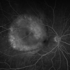
Late phase fluorescein angiogram of a 59-year-old female with a choroidal tumor secondary to metastatic breast cancer.
Photographer: David Saiz COT, Mayo Clinic Arizona
Imaging device: Optos California
Condition/keywords: breast cancer, FA late phase, metastatic cancer
-
 Breast cancer metastatic to choroid
Jul 13 2021 by Odette M. Houghton, MD
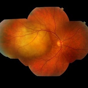
Montage photograph of a 59-year-old female with a choroidal tumor secondary to metastatic breast cancer.
Photographer: David Saiz COT, Mayo Clinic Arizona
Imaging device: Topcon
Condition/keywords: breast cancer, metastatic cancer, metastatic lesion
-
 Choroidal Metastasis With Orange Pigment in a Patient With Endometrial Carcinoma
Aug 8 2024 by Guilherme Sturzeneker, MD, MSc
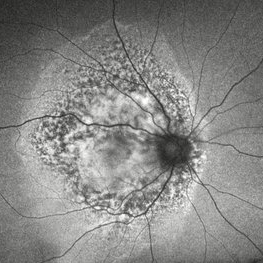
Ultra-widefield fundus photograph and autofluorescence of a 62-year-old woman with endometrial cancer, denoting choroidal metastasis with unusual orange pigment. This presentation is a reminder that the development of orange pigment is not pathognomonic for choroidal melanoma, as it may be seen in other lesions such as carcinoma metastasis.
Photographer: Andrea Almeida
Imaging device: Optos Silverstone
Condition/keywords: choroidal metastasis, metastatic cancer, orange pigment
-
 Choroidal Metastasis With Orange Pigment in a Patient With Endometrial Carcinoma
Aug 8 2024 by Guilherme Sturzeneker, MD, MSc
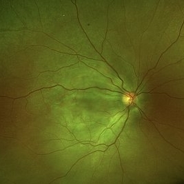
Ultra-widefield fundus photograph and autofluorescence of a 62-year-old woman with endometrial cancer, denoting choroidal metastasis with unusual orange pigment. This presentation is a reminder that the development of orange pigment is not pathognomonic for choroidal melanoma, as it may be seen in other lesions such as carcinoma metastasis.
Photographer: Andrea Almeida
Imaging device: Optos Silverstone
Condition/keywords: choroidal metastasis, metastatic cancer, orange pigment
-
 Ciliary Body Metastasis
Mar 26 2025 by Virginia Gebhart
54 year old female referred for iris mass. UBM shows large solid mass originating in the ciliary body and eroding into the anterior chamber under the iris epithelium. Recent CT scans revealed multiple bilateral pulmonary and hepatic nodules. Pt has been scheduled for PET scan and liver biopsy by radiation oncologist.
Photographer: Virginia Gebhart, Retina Consultants of Carolina
Imaging device: Samsung Galaxy
Condition/keywords: choroidal metastasis, ciliary body mass, metastatic cancer
-
 Extra-scleral Extension of Choroidal Melanoma
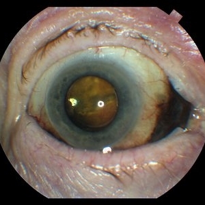
Dec 23 2021 by Jessica Norkus
89-year-old female with extra-scleral extension of choroidal metastatic melanoma. Patient hadn't been seen by any eye doctor in 3 years prior to this visit. Noticed sclera darkening about 6 months ago, with vision loss noted for about 4-5 months. Presented with LP vision. Emergent MRI of brain/orbit showed no extension beyond what is seen at slit lamp. CT C/A/P w/ contrast ordered and found 2 hepatic lesions, concerning for potential mets. Patient referred to medical oncology.
Photographer: Jessica Norkus, COA, OSC
Imaging device: Topcon TRC 50DX
Condition/keywords: extrascleral extension, metastatic cancer, metastatic lesion
-
 Extra-scleral Extension of Choroidal Melanoma
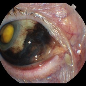
Dec 23 2021 by Jessica Norkus
89-yea- old female with extra-scleral extension of choroidal metastatic melanoma. Patient hadn't been seen by any eye doctor in 3 years prior to this visit. Noticed sclera darkening about 6 months ago, with vision loss noted for about 4-5 months. Presented with LP vision. Emergent MRI of brain/orbit showed no extension beyond what is seen at slit lamp. CT C/A/P w/ contrast ordered and found 2 hepatic lesions, concerning for potential mets. Patient referred to medical oncology.
Photographer: Jessica Norkus, COA, OSC
Imaging device: Topcon TRC 50DX
Condition/keywords: external photography, extrascleral extension, metastatic cancer, metastatic lesion
-
 Extra-scleral Extension of Choroidal Melanoma
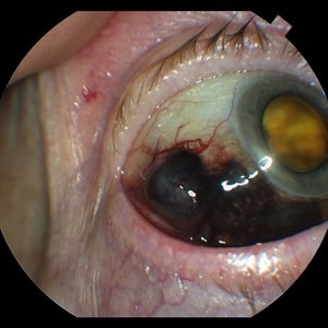
Dec 23 2021 by Jessica Norkus
89-year-old female with extra-scleral extension of choroidal metastatic melanoma. Patient hadn't been seen by any eye doctor in 3 years prior to this visit. Noticed scleral darkening about 6 months ago, with vision loss noted for about 4-5 months. Presented with LP vision. Emergent MRI of brain/orbit showed no extension beyond what is seen at slit lamp. CT C/A/P w/ contrast ordered and found 2 hepatic lesions, concerning for potential mets. Patient referred to medical oncology.
Photographer: Jessica Norkus, COA, OSC
Imaging device: Topcon TRC 50DX
Condition/keywords: external photography, extrascleral extension, metastatic cancer, metastatic lesion
-
 Extra-scleral Extension of Choroidal Melanoma
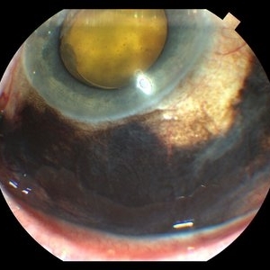
Dec 23 2021 by Jessica Norkus
89-year-old female with extra-scleral extension of choroidal metastatic melanoma. Patient hadn't been seen by any eye doctor in 3 years prior to this visit. Noticed scleral darkening about 6 months ago, with vision loss noted for about 4-5 months. Presented with LP vision. Emergent MRI of brain/orbit showed no extension beyond what is seen at slit lamp. CT C/A/P w/ contrast ordered and found 2 hepatic lesions, concerning for potential mets. Patient referred to medical oncology.
Photographer: Jessica Norkus, COA, OSC
Imaging device: Topcon TRC 50DX
Condition/keywords: extrascleral extension, metastatic cancer, metastatic lesion
-
 Extra-scleral Extension of Choroidal Melanoma
Dec 23 2021 by Jessica Norkus
89-year-old female with extra-scleral extension of choroidal metastatic melanoma. Patient hadn't been seen by any eye doctor in 3 years prior to this visit. Noticed scleral darkening about 6 months ago, with vision loss noted for about 4-5 months. Presented with LP vision. Emergent MRI of brain/orbit showed no extension beyond what is seen at slit lamp. CT C/A/P w/ contrast ordered and found 2 hepatic lesions, concerning for potential mets. Patient referred to medical oncology.
Photographer: Jessica Norkus, COA, OSC
Imaging device: Topcon TRC 50DX
Condition/keywords: external photography, extrascleral extension, metastatic cancer, metastatic lesion
-
 Metastatic Breast Carcinoma
Mar 26 2019 by Gary R. Cook, MD, FACS
64-year-old white female with metastatic breast carcinoma lesion superior to optic disc OD; VA= 20/70-1.
Imaging device: Topcon VT-50
Condition/keywords: breast cancer, breast carcinoma, choroidal metastasis, metastatic cancer, metastatic lesion
-
 Metastatic Cancer
Mar 26 2019 by Gary R. Cook, MD, FACS
64-year-old WF with metastatic breast carcinoma OD s/p radiation treatment; VA improved to 20/25.
Imaging device: Topcon VT-50
Condition/keywords: breast cancer, breast carcinoma, choroidal metastasis, metastatic lesion
-
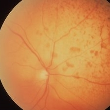 Metastatic Cancer
Mar 26 2019 by Gary R. Cook, MD, FACS
75-year-old white male with metastatic prostate cancer of the right eye; VA= counting fingers at 1 foot.
Imaging device: Topcon VT-50
Condition/keywords: choroidal metastasis, metastatic lesion, retinal hemorrhage
 Loading…