Search results (165 results)
-
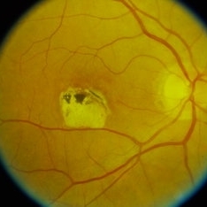
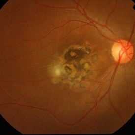
 ARMD with Large Macular Scar
Oct 12 2012 by Jeffrey G. Gross, MD, FASRS
ARMD with large macular scar in untreated subfoveal CNV.
Condition/keywords: choroidal neovascularization (CNV), macular scar, subfoveal choroidal neovascularization
-
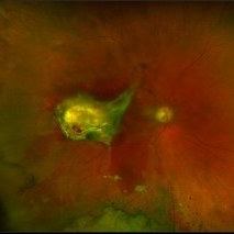
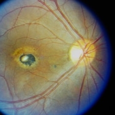
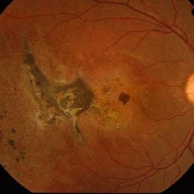
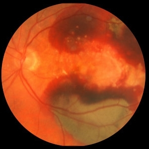 ARMD with Subretinal Hemorrhages and Macular Scarring
Oct 16 2012 by Jeffrey G. Gross, MD, FASRS
ARMD with subretinal hemorrhages and macular scarring, 20/400.
Condition/keywords: 20/400, macular scar, subretinal hemorrhage
-
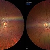
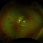 Chorioretinal Macula Scar (Ultrawide View)
May 12 2025 by Briana Hernandez
Ultra wide Optos image of Chorioretinal Macular Scar in 9-year-old female patient.
Photographer: Briana Hernandez, Hilton Head Retina Institute
Imaging device: Optos
Condition/keywords: chorioretinal scar, macular scar, ultra-wide field imaging
-
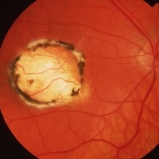
 Congenital Toxoplasmosis
Apr 8 2019 by Gary R. Cook, MD, FACS
Right eye of a 23-year-old male with congenital toxoplasmosis OD; view of macular lesion.
Condition/keywords: congenital toxoplasmosis, inactive toxoplasmosis, macular scar, ocular toxoplasmosis
-
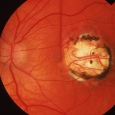
 Congenital Toxoplasmosis
Apr 8 2019 by Gary R. Cook, MD, FACS
23-year-old with congenital toxoplasmosis; view of optic disc and macular scar OS.
Condition/keywords: chorioretinal scar, congenital toxoplasmosis, inactive toxoplasmosis, macular scar, ocular toxoplasmosis
-
 Congenital Toxoplasmosis
Apr 8 2019 by Gary R. Cook, MD, FACS
Right eye of a 38-year-old female with bilateral congenital toxoplasmosis lesions; V.A. = 20/70 OD
Imaging device: Topcon VT-50
Condition/keywords: chorioretinal scar, congenital toxoplasmosis, inactive, inactive toxoplasmosis, macular scar, ocular toxoplasmosis
-
 Congenital Toxoplasmosis
Apr 8 2019 by Gary R. Cook, MD, FACS
Left eye of the same 38-year-old female with congenital toxoplasmosis lesion; V.A. = 20/40 due to temporal location of the Toxo scar.
Imaging device: Topcon VT-50
Condition/keywords: chorioretinal scar, congenital toxoplasmosis, inactive toxoplasmosis, macular scar, ocular toxoplasmosis
-
 Congenital Toxoplasmosis Macular Scarring
Nov 6 2021 by Emmanouil Gavalas, MD
Right eye OCT image showing atrophy and loss of foveal neuroretinal tissue and RPE.
Photographer: Emmanouil Gavalas MD, Ophthalmos Reseach and Educational Institute,Nicosia,Cyprus
Imaging device: Heidelberg Spectralis OCT
Condition/keywords: congenital toxoplasmosis, macular scar, ocular toxoplasmosis
-
 Congenital Toxoplasmosis Macular Scarring
Nov 6 2021 by Emmanouil Gavalas, MD
Fundus photographs of an 26-year-old female showing right eye macular scarring Incidental diagnosis Visual Acuity OD 6/12 OS 6/6
Photographer: Emmanouil Gavalas MD, Ophthalmos Reseach and Educational Institute,Nicosia,Cyprus
Imaging device: Zeiss Clarus 500
Condition/keywords: congenital toxoplasmosis, macular scar, ocular toxoplasmosis
-
 Congenital Toxoplasmosis Scar
Apr 8 2019 by Gary R. Cook, MD, FACS
5-year-old white male with a typical, deep, pigmented chorioretinal scar secondary to congenital toxoplasmosis OS.
Condition/keywords: chorioretinal scar, congenital toxoplasmosis, inactive toxoplasmosis, macular scar, ocular toxoplasmosis
-
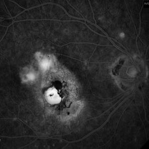
 cRORA
cRORA
Aug 5 2020 by Dhaivat Shah
A 54-year-old healthy male presented to us with a decreased vision in right eye since past 8 years. The patient gave a history of bleed in right eye before 8 years for which some intravitreal injection was given; post which there no major visual improvement. No details or documentation was available regarding the same. His BCVA in the right eye was 5/60. Fundus examination revealed a sharply demarcated hypopigmented patch over the macula with mild posterior excavation suggestive of macular scar. OCT image shows foveal thinning with loss of Retinal pigment epithelium and outer retinal layers (RORA). There are 2 types of RORAs, complete and incomplete. Complete RORA and incomplete RORA are entities defined by various imaging modalities describing atrophy of the retinal pigment epithelial and the outer retinal layers. OCT imaging defines incomplete RORA (iRORA) as a region of signal hyper transmission into the choroid and a corresponding zone of attenuation ordisruption of the RPE (<250um) and evidence of overlying photoreceptor degeneration (<250um). There should not be any RPE tear associated with it. OCT imaging describes complete RORA (cRORA) based on 4 inclusion criteria. These include, area of hypertransmission of more than 250um, zone of attenuation or disruption of the RPE of more than 250um in diameter, evidence of overlying photoreceptor degeneration and absence of scrolled RPE or other signs of an RPE tear. Other modalities used to define these include fundus autoflourescence(FAF), near infrared reflectance(NIR) and color fundus photograph(CFP). On CFP, it shows a sharply demarcated hypopigmented of >250um size with better visibility of choroidal vessels. FAF shows a hypo autoflourescent patch with sharply demarcated borders of size >250um, the colour of which is similar to that of the optic nerve head or retinal blood vessels excluding any pigmentation or artefact. On NIR, it shows a hyperreflective area with sharply demarcated borders of >250um size excluding any artefact. RORA can be seen in conditions like geographical atrophy in ARMD, central areolar choroidal dystrophy, atrophy secondary to anti-VEGF treatment. References: 1. Sadda SR, Guymer R, Holz FG, et al. Consensus Definition for Atrophy Associated with Age-Related Macular Degeneration on OCT: Classification of Atrophy Report 3 [published correction appears in Ophthalmology. 2019 Jan;126(1):177]. Ophthalmology. 2018;125(4):537-548. 2. Guymer RH, Rosenfeld PJ, Curcio CA, et al. Incomplete Retinal Pigment Epithelial and Outer Retinal Atrophy in Age-Related Macular Degeneration: Classification of Atrophy Meeting Report 4. Ophthalmology. 2020;127(3):394-409. 3. Eng VA, Rayess N, Nguyen HV, Leng T. Complete RPE and outer retinal atrophy in patients receiving anti-VEGF treatment for neovascular age-related macular degeneration. PLoS One. 2020;15(5):e0232353.
Photographer: Miss Anjum Zafar Khan
Imaging device: Choithram Netralaya
Condition/keywords: macular scar, outer retina, retinal pigment epithelium
-
 Dry Macular Scar With Recurrence
-
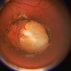
 Gunshot Injury
Dec 19 2024 by Angela Rico
53 y/o M who suffered gunshot wound to OD. Picture shows macular scar and sub retinal hemorrhage
Photographer: Angela Rico M.D.
Condition/keywords: macular scar, penetrating trauma
-
 IG
IG
May 17 2013 by Howard Schatz, MD
84-year-old white female, IG macular scar.
Condition/keywords: IG, macular scar
-
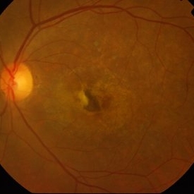
 Macular scar
Sep 21 2023 by Ben Serar
Fundus photograph of the RE showing scarring at the macula with hyperpigmention.
Condition/keywords: macular scar
-
 Macular scar
Sep 14 2023 by Ben Serar
Fundus photograph of RE showing hyper pigmented lesion at the posterior pole indicative of scarring at the macula.
Condition/keywords: macular scar
-
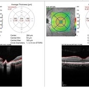
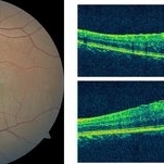 Macular Scar
Apr 7 2021 by Priya Rasipuram Chandrasekaran, MBBS, DO, DNB, FRCS
The fundus photo shows macular scar with epiretinal membrane in a 22-year-old female presenting with 20/80 N10 vision. The corresponding optical coherence tomography (vertical and horizontal raster scan) shows fibroglial proliferation with epiretinal membrane and traction at the fovea.
Condition/keywords: macular scar, scar
-
 Macular Scar
Jan 22 2018 by Jason Griffith
44-year-old male with history of CR scarring secondary to histoplasmosis.
Photographer: Jason Griffith, Tennessee Retina, Nashville TN
Imaging device: Topcon TRC 50EX
Condition/keywords: macular scar
-
 Macular Scar
Apr 12 2014 by Dipankar Barua, M.Sc
The 31-year-old male patient with visual acuity of right eye is 6/6 and 6/60 in left eye.
Photographer: Dipankar Barua
Imaging device: Topcon TRC 50 DX (IA)
Condition/keywords: macula, macular scar
-
 Macular Scar
Apr 14 2014 by Dipankar Barua, M.Sc
Female patient, 25-years-old. On examination, her vision of the right eye is 6/60 and left eye is 6/6. It seems to be a case of macular scar.
Photographer: Dipankar Barua
Imaging device: Topcon TRC 50 DX (IA)
Condition/keywords: macular scar
-
 Macular Scar
Apr 16 2014 by Dipankar Barua, M.Sc
Male patient, 15-years-old. Vision of the right eye is counting fingers and left eye is normal. It seems to be a case of macular scar in right eye.
Photographer: Dipankar Barua
Imaging device: Topcon TRC 50 DX (IA)
Condition/keywords: macular scar
-
 Macular Scar
Apr 16 2014 by Dipankar Barua, M.Sc
Male patient, 15-years-old. Vision of the right eye is counting fingers and left eye is normal. It seems to be a case of Macular scar in right eye.
Photographer: Dipankar Barua
Imaging device: Topcon TRC 50 DX (IA)
Condition/keywords: macular scar
-
 Macular Scar
Apr 16 2014 by Dipankar Barua, M.Sc
Male patient, 15-years-old. Vision of the right eye is counting fingers and left eye is normal. It seems to be a case of macular scar in right eye.
Photographer: Dipankar Barua
Imaging device: Topcon TRC 50 DX (IA)
Condition/keywords: macular scar
-
 Macular Scar
Apr 18 2014 by Dipankar Barua, M.Sc
Female patient, 55-years-old. On examination her vision in the right eye is 6/9 and left eye is only hand movement. It seems to be a case of macular scar in left eye.
Photographer: Dipankar Barua
Imaging device: Topcon TRC 50 DX (IA)
Condition/keywords: macular scar
 Loading…