Search results (6 results)
-
 Kyrieleis Plaques
Jan 12 2022 by Joshua S Agranat, MD
Prominent kyrieleis plaques in a patient with toxoplasmosis.
Condition/keywords: Kyrieleis arteritis, kyrieleis plaques, toxoplasmosis
-
 Kyrieleis Plaques
Aug 18 2025 by Helder Vasconcelos
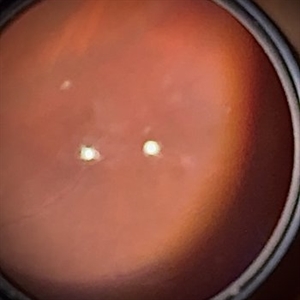
Kyrieleis Plaques and vitritis in a patient with posterior uveitis secondary to presumed ocular toxoplasmosis.
Photographer: Helder Vasconcelos
Imaging device: Smartphone Fundoscopy
Condition/keywords: kyrieleis plaques, toxoplasmosis chorioretinitis
-
 Occlusive Retinal Vasculitis
Oct 3 2024 by Logan ryzenga
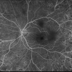
4 view ultra-widefield Optos fluorescein angiogram in the left eye of a 39 year old woman occlusive retinal vasculitis with four quadrant Kyrieleis plaques. This is a showcase of a suspected, rarely reported, and atypical presentation of Behcet's Syndrome.
Photographer: Logan Ryzenga
Imaging device: Optos California
Condition/keywords: Behcet's Disease, Behcet's uveitis, Fluorescein angiography, fluorescein leakage, kyrieleis plaques, non-perfusion, OPTOS, OPTOS CALIFORNIA, ultra-wide field imaging, Uveitis
-
 Retinal Vasculitis
Mar 26 2025 by Korey Starkey
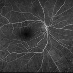
41 year-old patient presents with vascular FA findings of occlusive vasculitis with four quadrant Kyrieleis plaques OU showcases a possibly rare but reported atypical presentation of Behcet's Syndrome.
Photographer: Korey Starkey
Imaging device: Optos
Condition/keywords: Behcet's Disease, FA early phase, Fundus Fluorescein Angiography, Optos, retinal vasculitis, ultra-wide field imaging, venous beading
-
 Retinal Vasculitis
Mar 26 2025 by Korey Starkey
41 year-old patient presents with vascular FA findings of occlusive vasculitis with four quadrant Kyrieleis plaques OU showcases a possibly rare but reported atypical presentation of Behcet's Syndrome.
Photographer: Korey Starkey
Imaging device: Optos
Condition/keywords: FA early phase, Fundus Fluorescein Angiography, ischemia, Optos, retinal vasculitis, ultra-wide field imaging, venous beading
-
 Toxoplasmosis Slide 2
Oct 22 2012 by Ronald C. Gentile, MD
One month following treatment with Bactrim, Clindamycin, and oral prednisone the focal area chorioretinitis has coalesced with a decrease in overlying vitreous inflammation. Kyrieleis plaques can be seen along the inferior retinal arteriole.
Photographer: The New York Eye & Ear Infirmary Department of Medical Imaging
Condition/keywords: posterior uveitis, toxoplasmosis
 Loading…