Search results (11 results)
-
 Iris Melanoma
-
 Iris Melanoma
Jul 11 2013 by Jason S. Calhoun
Iris melanoma.
Photographer: Jason S. Calhoun, Department of Ophthalmology, Mayo Clinic Jacksonville, Florida
Condition/keywords: iris melanoma
-
 Iris Melanoma
Jul 11 2013 by Jason S. Calhoun
Iris melanoma.
Photographer: Jason S. Calhoun, Department of Ophthalmology, Mayo Clinic Jacksonville, Florida
Condition/keywords: iris melanoma
-
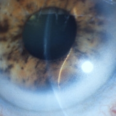
 Iris Melanoma
May 19 2017 by Nichole Lewis
Iris melanoma.
Photographer: Nichole Lewis
Condition/keywords: iris melanoma
-
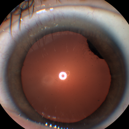
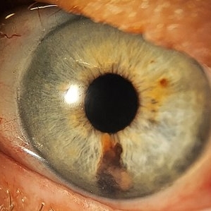
 Iris Melanoma
Jan 28 2025 by Korey Starkey
Slit-lamp image of 90-year-old patient with iris melanoma and new hemorrhage affecting the right eye. Patient re-presented after nearly 1 year, now seeking treatment. Given iris location of tumor, multiple clock hours of iris involved, and increase in size of the known malignant transformation; safest approach was enucleation.
Photographer: Korey Starkey
Imaging device: Slit lamp camera
Condition/keywords: anterior chamber, hemorrhage, iris melanoma, slit lamp photo
-
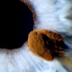
 Iris Melanoma
Feb 1 2024 by Virginia Gebhart
90 year old female with elevated pigmented lesion, amelanotic portion extending toward the angle, questionable vascularity on UBM.
Photographer: Virginia Gebhart
Imaging device: Samsung Galaxy Z Flip
Condition/keywords: iris lesion, iris melanoma
-
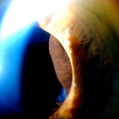
 New Iris Melanoma
Oct 10 2024 by Virginia Gebhart
56 year old male with new amelanotic melanoma emanating from the ciliary body through the posterior iris epithelium. CT scan showed no evidence of metastatic disease. Pt scheduled for radioactive plaque and tumor biopsy
Photographer: Virginia Gebhart, Retina Consultants of Carolina
Imaging device: Samsung Galaxy
Condition/keywords: amelanotic melanoma, iris melanoma
-
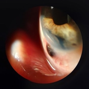 Scleral Ectasia Post Radiation for Iris Melanoma
Jul 5 2024 by Zach Seim
Slit-Lamp Photograph of a 52 year old male with Scleral Ectasia post radiation for Iris Melanoma.
Photographer: Zach Seim
Imaging device: Slit Lamp Photography on Samsung Galaxy 7
Condition/keywords: Iris, iris melanoma, scleral ectasia, slit lamp photo, slit lamp photography
-
 Slide 14-30
Mar 4 2019 by Lancaster Course in Ophthalmology
The vast majority of iris melanomas are composed of spindle A and/or spindle B cells and are relatively benign.
Condition/keywords: iris melanoma, melanoma, spindle A melanoma, spindle B melanoma, spindle cells
-
 Slide 14-31
Mar 4 2019 by Lancaster Course in Ophthalmology
The vast majority of iris melanomas are composed of spindle A and/or spindle B cells and are relatively benign.
Condition/keywords: iris melanoma, melanoma, spindle A melanoma, spindle B melanoma, spindle cells
-
 Suspicious Lesion 18 Years s/p Iris Resection
Oct 15 2024 by Virginia Gebhart
85 year old female with small pigmented lesion present s/p sectoral iridectomy in 2006. Lesion is suspicious for recurrence of melanoma after 18 years. Stable compared to previous exam in March 2024, unclear if this is a new lesion or has been present for an extended time. Will monitor closely.
Photographer: Virginia Gebhart, Retina Consultants of Carolina
Imaging device: Samsung Galaxy
Condition/keywords: iris melanoma, melanoma
 Loading…