Search results (73 results)
-
 Broken macroaneurysm
Nov 27 2022 by Nassim Alejandro Abreu Arbaje, MD
Fundus video frame of a 58 year old male who had a PPV on his left eye because a retinal macroaneurysm that broke a bled on all 3 retinal planes.
Photographer: Nassim Abreu, Hospital Dr. Elías Santana
Imaging device: NGenuity 3D system
Condition/keywords: broken macroanerysm, intraretinal hemorrhage, macroaneurysm, subretinal hemorrhage, vitreous hemorrhage
-
 BRVO With Decreased Vision from Ruptured Aneurysm in Area of Collateral Vessels
Dec 27 2017 by John S. King, MD
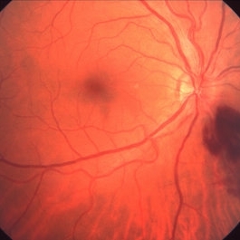
BRVO with decreased vision from ruptured aneurysm in area of collateral vessels; central IRH.
Imaging device: Cirrus
Condition/keywords: arteriolar macroaneurysm, branch retinal vein occlusion (BRVO), collateral retinal vessel, intraretinal hemorrhage
-
 Chronic Retinal Vein Occlusion
Jul 8 2012 by Jeffrey S. Heier, MD
Chronic RVO with vascular changes, intraretinal hemorrhages
Imaging device: Zeiss
Condition/keywords: chronic retinal vein occlusion, intraretinal hemorrhage
-
---thumb.jpg/image-square;max$300,300.ImageHandler) Fibrovascular Proliferation
Fibrovascular Proliferation
Feb 13 2013 by From the Collections of Thomas M. Aaberg, MD and Thomas M. Aaberg Jr., MD
Neovascularization, fibrous proliferation, intraretinal hemorrhage.
Condition/keywords: fibrous proliferation, intraretinal hemorrhage, neovascularization (NV)
-
 Idiophatic retinal vasculitis
Jul 9 2023 by Luiz A Zago, PhD
Idiophatic retinal vasculitis in a 45 year old woman
Photographer: Luiz Zago, PhD.
Imaging device: Topcon 50IX
Condition/keywords: diffuse vasculitis, intraretinal hemorrhage, optic disc leakage
-
 Idiophatic vasculitis OD
Jul 9 2023 by Luiz A Zago, PhD
Idiophatic vasculitis
Photographer: Luiz Zago, PhD.
Imaging device: Topcon 50IX
Condition/keywords: intraretinal hemorrhage, leaky parafoveal capillaries, optic disc leakage, vasculitis
-
 Inferonasal Branch Retinal Vein Occlusion
Aug 23 2012 by Gerardo Garcia-Aguirre, MD
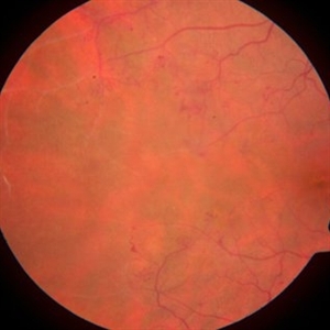
Fundus of a 55-year-old male showing intraretinal hemorrhages in the inferonasal quadrant.
Photographer: Noemí Hernández, Asociación para Evitar la Ceguera en México
Condition/keywords: branch retinal vein occlusion (BRVO), intraretinal hemorrhage
-
 Inferonasal BRVO - Fluorescein Angiogram
Aug 23 2012 by Gerardo Garcia-Aguirre, MD
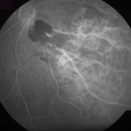
Fluorescein angiogram showing hypofluorescence secondary to intraretinal hemorrhages, and perivascular hyperfluorescence secondary to vascular incompetence.
Photographer: Noemí Hernández, Asociación para Evitar la Ceguera en México
Condition/keywords: branch retinal vein occlusion (BRVO), intraretinal hemorrhage, vascular incompetence
-
---thumb.jpg/image-square;max$300,300.ImageHandler) Intraretinal Hemorrhage
Intraretinal Hemorrhage
Feb 15 2013 by From the Collections of Thomas M. Aaberg, MD and Thomas M. Aaberg Jr., MD
Color fundus photograph showing vascular sheathing and diffuse intraretinal hemorrhage associated with presumed viral retinitis.
Condition/keywords: retinal hemorrhage
-
---thumb.jpg/image-square;max$300,300.ImageHandler) Intraretinal Hemorrhage 4
Intraretinal Hemorrhage 4
Mar 20 2013 by Maurice F. Rabb
62-year- old white male with a small hemorrhage in the posterior segment in the right eye. In the left eye, the superior portion of the fundus was a uniform creamy white color with appearance of closure of the superior temporal artery. There were several areas of intraretinal hemorrhage and whitish lesions associated with the hemorrhage. There was vitreous haze, more marked inferiorly.
Condition/keywords: closure of superior temporal artery, intraretinal hemorrhage, vitreous haze, whitish lesions
-
---thumb.jpg/image-square;max$300,300.ImageHandler) Intraretinal Hemorrhage 1
Intraretinal Hemorrhage 1
Mar 20 2013 by Maurice F. Rabb
62-year- old white male with a small hemorrhage in the posterior segment in the right eye. In the left eye, the superior portion of the fundus was a uniform creamy white color with appearance of closure of the superior temporal artery. There were several areas of intraretinal hemorrhage and whitish lesions associated with the hemorrhage. There was vitreous haze, more marked inferiorly.
Condition/keywords: closure of superior temporal artery, intraretinal hemorrhage, vitreous haze, whitish lesions
-
---thumb.jpg/image-square;max$300,300.ImageHandler) Intraretinal Hemorrhage 2
Intraretinal Hemorrhage 2
Mar 20 2013 by Maurice F. Rabb
62-year- old white male with a small hemorrhage in the posterior segment in the right eye. In the left eye, the superior portion of the fundus was a uniform creamy white color with appearance of closure of the superior temporal artery. There were several areas of intraretinal hemorrhage and whitish lesions associated with the hemorrhage. There was vitreous haze, more marked inferiorly.
Condition/keywords: closure of superior temporal artery, intraretinal hemorrhage, vitreous haze, whitish lesions
-
---thumb.jpg/image-square;max$300,300.ImageHandler) Intraretinal Hemorrhage 3
Intraretinal Hemorrhage 3
Mar 20 2013 by Maurice F. Rabb
62-year- old white male with a small hemorrhage in the posterior segment in the right eye. In the left eye, the superior portion of the fundus was a uniform creamy white color with appearance of closure of the superior temporal artery. There were several areas of intraretinal hemorrhage and whitish lesions associated with the hemorrhage. There was vitreous haze, more marked inferiorly.
Condition/keywords: closure of superior temporal artery, intraretinal hemorrhage, vitreous haze, whitish lesions
-
---thumb.jpg/image-square;max$300,300.ImageHandler) Intraretinal Hemorrhages (Case 2)
Intraretinal Hemorrhages (Case 2)
-
---thumb.jpg/image-square;max$300,300.ImageHandler) Intraretinal Hemorrhages (Case 2)
Intraretinal Hemorrhages (Case 2)
-
---thumb.jpg/image-square;max$300,300.ImageHandler) Intraretinal Hemorrhages (Case 2)
Intraretinal Hemorrhages (Case 2)
-
 Lyme Disease
Feb 13 2013 by From the Collections of Thomas M. Aaberg, MD and Thomas M. Aaberg Jr., MD
Papilledema, intra-retinal hemorrhage, periopticneuritis.
Condition/keywords: intraretinal hemorrhage, Lyme disease, periopticneuritis
-
---thumb.jpg/image-square;max$300,300.ImageHandler) Optic Disc and Retinal Edema
Optic Disc and Retinal Edema
Feb 13 2013 by From the Collections of Thomas M. Aaberg, MD and Thomas M. Aaberg Jr., MD
Intra-retinal hemorrhage papilledema.
Condition/keywords: intraretinal hemorrhage, optic disc, papilledema, retinal edema
-
---thumb.jpg/image-square;max$300,300.ImageHandler) Peripapillary Atrophy
Peripapillary Atrophy
Feb 13 2013 by From the Collections of Thomas M. Aaberg, MD and Thomas M. Aaberg Jr., MD
Papilledema, intra-retinal hemorrhage, periopticneuritis.
Condition/keywords: intraretinal hemorrhage, papilledema, periopticneuritis, peripapillary atrophy
-
 Progressive Outer Retinal Necrosis
Nov 30 2018 by Nichole Lewis
Fluorescein angiogram of an 86-year-old male with progressive outer retinal necrosis and chronic cystoid macular edema. This patient has occlusive vasculitis with non-perfusion, significant retinitis, retinal whitening and intra-retinal hemorrhages. Patient is immunosupressed with a history of kidney transplantation. VA 20/60. Patient was treated with intravitreal foscarnet and admitted to the hospital for an infectious disease and transplant team consultation.
Photographer: Nichole Lewis
Condition/keywords: cystoid macular edema (CME), intraretinal hemorrhage, non-perfusion, occlusive vasculitis, progressive outer retinal necrosis (PORN), retinal whitening, retinitis
-
 Progressive Outer Retinal Necrosis
Nov 5 2019 by Nichole Lewis
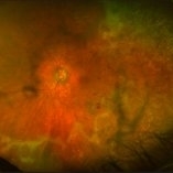
86-year-old male with progressive outer retinal necrosis, significant retinitis, retinal whitening, intraretinal hemorrhages and peripheral rpe changes. FA showed occlusive vasculitis with non-perfusion. Patient is immuno-suppressed with a history of renal transplant. VA 20/60.
Photographer: Nichole Lewis
Imaging device: Optos
Condition/keywords: intraretinal hemorrhage, occlusive vasculitis, progressive outer retinal necrosis (PORN), retinal pigment epithelium (RPE) changes, retinal whitening, retinitis
-
 Ruptured Macroaneurysm
May 22 2019 by Nichole Lewis
FA of a 91-year-old woman with a ruptured macroaneurysm, intraretinal hemorrhage and subretinal hemorrhage. VA 20/400.
Photographer: Nichole Lewis
Condition/keywords: intraretinal hemorrhage, ruptured macroaneurysm, subretinal hemorrhage
-
 Intraretinal hemorrhage - post treatment
Feb 15 2013 by From the Collections of Thomas M. Aaberg, MD and Thomas M. Aaberg Jr., MD
Color fundus photograph of the eye after systemic treatment showing significant reduction in intraretinal hemorrhage.
Condition/keywords: retinal hemorrhage
-
---thumb.jpg/image-square;max$300,300.ImageHandler) Intraretinal Hemorrhage and Premacular Fibrosis - post treatment
Intraretinal Hemorrhage and Premacular Fibrosis - post treatment
Feb 15 2013 by From the Collections of Thomas M. Aaberg, MD and Thomas M. Aaberg Jr., MD
Color fundus photograph of eye after systemic treatment showing significant reduction in intraretinal hemorrhage and premacular fibrosis.
Condition/keywords: macular pucker, retinal hemorrhage
-
---thumb.jpg/image-square;max$300,300.ImageHandler) intraretinal hemorrhage and retinal exudation
intraretinal hemorrhage and retinal exudation
Feb 15 2013 by From the Collections of Thomas M. Aaberg, MD and Thomas M. Aaberg Jr., MD
intraretinal hemorrhage and retinal exudation arising from microvascular damage associated with presumed herpesvirus infection.
Condition/keywords: macular edema, microangiopathy, retinal necrosis
 Loading…