Search results (73 results)
-
 Central Serous Retinopathy
Mar 19 2024 by Corey Grant
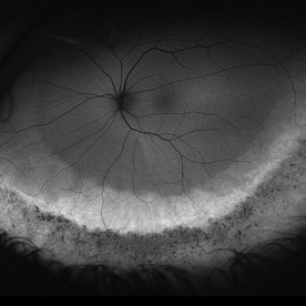
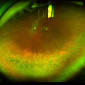
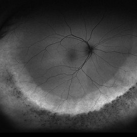
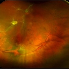
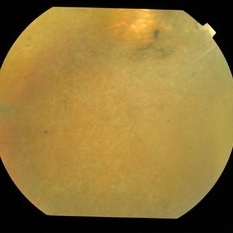
Ultra Wide-Field Fundus Autofluorescence Imaging of a 37 year old female with Central Serous Retinopathy affecting her right eye. Patient Visual Acuity was 20/20 in both eyes. Patient reported black spots in her vision onset three years ago, with associating flashes of light. Patient reports history of cortisone back injections a few years ago and denies Flonase use. The physician stated that there is hyperautofluorescence in the area of gutter of Sub-Retinal Fluid which likely happened from CSR.
Photographer: Corey Grant, OSC
Imaging device: OPTOS CALIFORNIA RGB
Condition/keywords: Central Serous Chorioretinopathy (CSR), central serous retinopathy (CSR), fundus autofluorescence (FAF), Guttering, hyperautofluorescence, inferior retina, OPTOS, Retina, Right Eye, subretinal fluid, ULTRA WIDE FIELD
-
 Coats' Disease
Jul 16 2019 by Kim Barrett
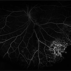
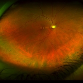
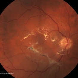
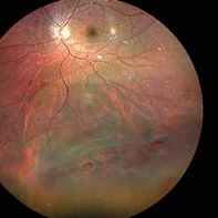
Ultra-wide field fluorescein angiogram of a 23-year-old male with Coats' disease, presented with distorted vision affecting his left eye. He reported seeing flashes and floaters since January of 2019, but the flashes had resolved. He was treated with Intravitreal Preservative Free Triamcinolone in the office and scheduled for PRP laser in the near future.
Photographer: Kim Barrett
Imaging device: Optos
Condition/keywords: Coats' disease, fluorescein angiogram (FA), fluorescein leakage, inferior retina, ischemia, left eye, Optos, ultra-wide field imaging
-
 Dislocated Crystalline Lens
Mar 19 2024 by Annaka Gooding
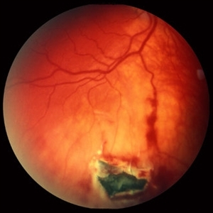
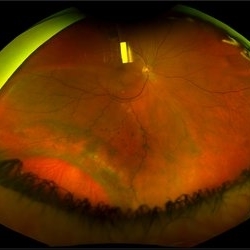
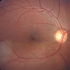
Ultra Wide field fundus photography of a 70 year old male who presented to clinic with a sudden increase of vision due to dropped crystalline lens secondary to severely dense cataract. Patient reported seeing a full black circle in his inferior visual field. Patient's visual acuity at time of visit was 20/100 with a +5.00 diopter lens. The physician recommended surgical intervention, and discussed surgery for PPV/PPL/IOL implantation with an ACIOL.
Photographer: Annaka Gooding, CPO
Imaging device: Optos California RGB
Condition/keywords: dislocated crystalline lens, fundus photography, inferior retina, OPTOS CALIFORNIA RGB, Right Eye, Ultra-wide field retinal imaging
-
 Exudative Retinal Detachment and Branch Retinal Vein Occulsion
Oct 29 2020 by Olivia Rainey
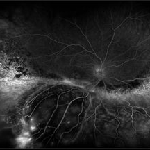
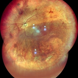
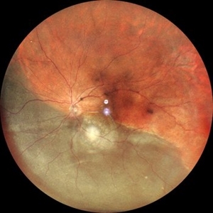
Ultra-widefield fluorescein anigogram of a 51-year-old female with an exudative retinal detachment and branch retinal vein occlusion with retinal neovascularization affecting her right eye. The physician stated that the multiple aneurysmal dilations noted in the inferior periphery are responsible for the exudative RD seen on exam. He is considering Coat's vs FEVR given family history of aneurysms/congenital heart pathology per patient. He encouraged the patient to control their blood pressure, cholesterol, blood sugar, and co-morbidities which may have promoted the BRVO. He recommended antiVEGF injections to control the vascular leakage. Given the severe presentation and imminent threat to her vision, he recommended Eylea as first line therapy.
Photographer: Olivia Rainey, OCT-C, COA
Imaging device: Optos California
Condition/keywords: branch retinal vein occlusion (BRVO), chronic retinal detachment, fluorescein angiogram (FA), fluorescein leakage, inferior retina, inferior retinal detachment, Optos, ultra-wide field imaging
-
 Intraocular Foreign Body, Metallic, in Inferior Retina with Hemorrhage
Oct 1 2012 by Jeffrey G. Gross, MD, FASRS
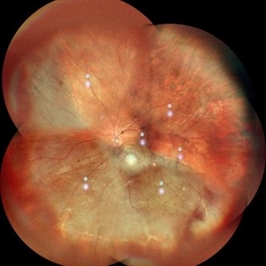
IOFB, metallic, in inferior retina with hemorrhage.
Condition/keywords: inferior retina, intraocular foreign body
-
 Ozurdex Implant Related Tear
Jan 26 2022 by Tracey Grabowski
Ultra wide-field photograph of a 73-year-old female with an Ozurdex implant causing a retinal tear in the inferior retina. Prompt laser was added to prevent a retinal detachment and patient has been doing well since. Patient had no symptoms following the occurrence.
Photographer: Tracey Grabowski
Imaging device: Optos California
Condition/keywords: fundus photograph, inferior retina, optos, ozurdex, Ozurdex implant, retinal tear, treated retinal tear, ULTRA WIDE FIELD
-
 Pigmentary Retinal Dystrophy
May 5 2020 by Olivia Rainey
Ultra-widefield fundus autofluorescence image of an 44-year-old male with pigmentary retinal dystrophy affecting both eyes. He presented with decreased night vision for 6 months prior to his appointment. He stated that his recovery time from transitioning from dark to light areas is reduced. He stated that his peripheral vision has never been very good for most of his life. He admits to environmental hearing loss. Patient denies family history of blin. His vision was 20/20 in both eyes. His full field ERG, visual fields were not consistent with RP. Genetic testing with ID Your IRD and annual follow up has been recommended.
Photographer: Olivia Rainey, OCT-C, COA
Imaging device: Optos California
Condition/keywords: fundus autofluorescence (FAF), hyperautofluorescence, hypoautofluorescence, inferior retina, left eye, Optos, ultra-wide field imaging
-
 Pigmentary Retinal Dystrophy
May 5 2020 by Olivia Rainey
Ultra-widefield pseudocolor image of an 44-year-old male with pigmentary retinal dystrophy affecting both eyes. He presented with decreased night vision for 6 months prior to his appointment. He stated that his recovery time from transitioning from dark to light areas is reduced. He stated that his peripheral vision has never been very good for most of his life. He admits to environmental hearing loss. Patient denies family history of blin. His vision was 20/20 in both eyes. His full field ERG, visual fields were not consistent with RP. Genetic testing with ID Your IRD and annual follow up has been recommended.
Photographer: Olivia Rainey, OCT-C, COA
Imaging device: Optos California
Condition/keywords: inferior retina, left eye, Optos, pigment, pseudocolor, ultra-wide field imaging
-
 Pigmentary Retinal Dystrophy
May 5 2020 by Olivia Rainey
Ultra-widefield fundus autofluorescence image of an 44-year-old male with pigmentary retinal dystrophy affecting both eyes. He presented with decreased night vision for 6 months prior to his appointment. He stated that his recovery time from transitioning from dark to light areas is reduced. He stated that his peripheral vision has never been very good for most of his life. He admits to environmental hearing loss. Patient denies family history of blin. His vision was 20/20 in both eyes. His full field ERG, visual fields were not consistent with RP. Genetic testing with ID Your IRD and annual follow up has been recommended.
Photographer: Olivia Rainey, OCT-C, COA
Imaging device: Optos California
Condition/keywords: fundus autofluorescence (FAF), hyperautofluorescence, hypoautofluorescence, inferior retina, Optos, pigment, ultra-wide field imaging
-
 Pigmentary Retinal Dystrophy
May 5 2020 by Olivia Rainey
Ultra-widefield pseudocolor image of an 44-year-old male with pigmentary retinal dystrophy affecting both eyes. He presented with decreased night vision for 6 months prior to his appointment. He stated that his recovery time from transitioning from dark to light areas is reduced. He stated that his peripheral vision has never been very good for most of his life. He admits to environmental hearing loss. Patient denies family history of blin. His vision was 20/20 in both eyes. His full field ERG, visual fields were not consistent with RP. Genetic testing with ID Your IRD and annual follow up has been recommended.
Photographer: Olivia Rainey, OCT-C, COA
Imaging device: Optos California
Condition/keywords: inferior retina, Optos, pigmentary retinal dystrophy, pseudocolor, ultra-wide field imaging
-
 Retinal Detachment with Giant Tear
Mar 13 2018 by Olivia Rainey
Ultra-wide field pseduocolor image of a 36-year-old male with an giant inferior tear, causing a retinal detachment.
Photographer: Olivia Rainey
Imaging device: Optos
Condition/keywords: color fundus photograph, giant retinal tear, inferior retina, macular splitting, Optos, ultra-wide field imaging
-
 Slide 4-3
Feb 20 2019 by Lancaster Course in Ophthalmology
Juvenile retinoschisis, a disorder with a sex-linked recessive pattern of inheritance. Clinical picture showing schisis in the inferior retina.
Condition/keywords: inferior retina, juvenile retinoschisis
-
 Tractional Retinal Detachment
Aug 22 2019 by Stacie Neview
Ultra-wide field pseudo-color photograph of 47-year-old male with diabetic retinopathy and subsequent diabetic tractional detachment. Patient was lost to follow up for 9 months after receiving anti-VEGF injections with mild PRP and presented with blurry vision.
Photographer: Stacie Neview
Imaging device: Optos
Condition/keywords: diabetic macular edema, diabetic retinopathy, inferior retina, left eye, pan-retinal photocoagulation (PRP), proliferative diabetic retinopathy (PDR), tractional retinal detachment
-
 Inferior RD OD with Exposed Scleral Buckle OD in Previous Images
Feb 4 2013 by James B. Soque, CRA, OCT-C, COA, FOPS
Fundus image of 66-year-old WM with Hx of SBOD in 2009. presents with exposed SBOD and infection seen in accompanying images.
Photographer: James Soque, CRA COA
Imaging device: Topcon TRC 50 DX, MERGE Imaging Software
Condition/keywords: inferior retinal detachment
-
 Inferior retinal detachment
Dec 19 2012 by Eric A. Postel, MD
Color fundus photograph of an inferior retinal detachment
-
 Inferior Retinal Detachment with Lattice
Sep 30 2020 by Sham Talati, DOMS
A patient of inferior retinal detachment with lattice inferiorly.
Photographer: Dr. Sham Talati,Retina Foundation,Ahmedabad
Imaging device: Nidek Mirante
Condition/keywords: lattice degeneration
-
 Inferior retinal detachment with lattice and holes
May 31 2023 by Aditya S Kelkar, MS, FRCS, FASRS,FRCOphth
Importance of dilated retina check up before Lasik surgery can't be better demonstrated...patient totally asymptomatic came for Lasik opinion and has inferior retinal detachment with lattice and holes, sparing the macula
Photographer: Dr. Sahil Wagh , National Institute of Opthalmology, Pune , India
Imaging device: Zeiss Clarus 500
Condition/keywords: inferior retinal detachment
-
 Retinal Detachment
May 13 2016 by Nichole Lewis
Inferior Retinal Detachment with some demarcation line s/p barrier laser.
Photographer: Nichole Lewis
Condition/keywords: barrier laser
-
 Retinal Detachment
Nov 3 2023 by Anjana Mirajkar, MS Ophthalmology
A widefield image of OS of a 55 year old female case of inferior retinal detachment with macula off.
Photographer: Dr. Anjana Mirajkar -Retina Foundation, Ahmedabad
Imaging device: Mirante-Nidek
Condition/keywords: inferior retinal detachment, Retinal Detachment
-
 Retinal Detachment
Nov 3 2023 by Anjana Mirajkar, MS Ophthalmology
A widefield image (montage) of OS of a 55 year old female case of inferior retinal detachment with macula off.
Photographer: Dr. Anjana Mirajkar -Retina Foundation, Ahmedabad
Imaging device: Mirante-Nidek
Condition/keywords: inferior retinal detachment, Retinal Detachment
-
 Retinal Detachment After Retinoblastoma Treatment
Mar 10 2024 by Alexandre Grandinetti, MD, PhD
Inferior retinal detachment occurring 6 years after treatment with intraarterial chemotherapy and laser in an 8-year-old boy.
Photographer: Corina Szrek
Condition/keywords: pediatric, retinoblastoma
-
 Retinitis Pigmentosa
Sep 22 2014 by Mallika Goyal, MD
Inferior retina of a 32-year-old lady with bilateral retinitis pigmentosa. She has progressive visual complaints starting at age 5, and is the offspring of a consanguineous marriage. Marked disc pallor, retinal arteriolar attenuation, pigment disturbance and macular degeneration are classic features.
Photographer: Mallika Goyal, MD, Apollo Health City, Jubilee Hills, Hyderabad-500033
Condition/keywords: retinitis pigmentosa
-
 Slide 9-73
Feb 26 2019 by Lancaster Course in Ophthalmology
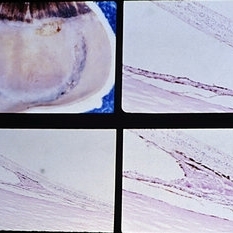
Inferior retinal dialysis with a localized area of long-standing retinal detachment and demarcation line (upper left). There is total atrophy of the photoreceptor cell layer (upper right). The demarcation line (lower views) is an area of RPE hypertrophy and hyperplasia with nodular basement membrane production and retinal adhesion.
Condition/keywords: photoreceptor cell, retinal dialysis, retinal pigment epithelium (RPE) hypertrophy
-
 Vasoproliferative Tumor
Aug 29 2024 by César Adrián Gómez Valdivia, MD
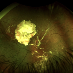
Inferior retinal vasoproliferative tumor found in a 66 year-old female patient. Asymptomatic.
Photographer: @eyemissu2
Imaging device: California ICG OPTOS
Condition/keywords: Vasoproliferative Tumor
-
 Acute Exudative Polymorphous Vitelliform Maculopathy Angio OD
Aug 27 2014 by Flavio A. Rezende, MD, PhD
45-year-old man with mild decrease in vision after strong headache. Fundus showing multiple deep irregular vitelliform lesions spread throughout entire posterior pole OU, forming a typical level of subretinal confluent lesions at the inferior retinal vascular arcades. No primary tumor or metastasis found.
Photographer: Eduardo Martins, Pontifícia Universidade Católica - Rio de Janeiro, Brazil
Imaging device: Topcon TRC 50EX
Condition/keywords: polymorphous exudative vitelliform maculopathy
 Loading…