Search results (11 results)
-
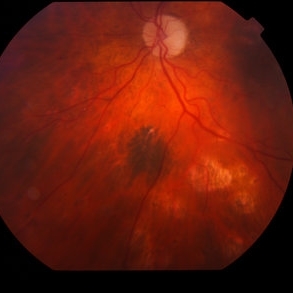
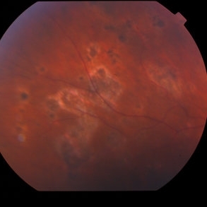
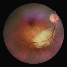 Ocular toxoplasmosis
Mar 5 2023 by Sergio Emilio Sifuentes Renteria, MD
Color fundus photograph of the right eye of a patient with HIV-infection and concomitant ocular toxoplasmosis.
Photographer: Sergio Emilio Sifuentes Rentería - Clínica Especializada Condesa Iztapalapa
Condition/keywords: HIV, infectious uveitis, posterior uveitis, toxoplasmosis, toxoplasmosis chorioretinitis
-
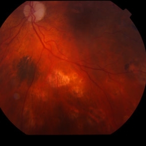
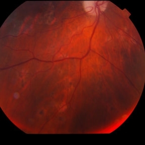
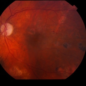 West Nile Virus Choroiditis
Apr 4 2014 by Suber S. Huang, MD, MBA, FASRS
Fundus photograph (11 month follow-up) of an 88-year-old woman who developed West Nile virus encephalitis on 08/2012 and subsequent choroiditis.
Photographer: Geoffrey Pankhurst; University Hospitals Eye Institute, Case Western Reserve University, Cleveland, OH
Imaging device: TopCon TRC50EX
Condition/keywords: choroiditis, disseminated choroiditis, infectious uveitis, optic nerve atrophy
-
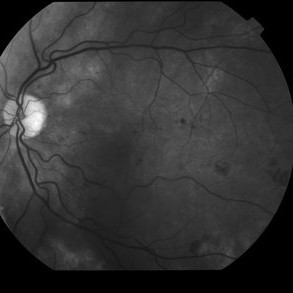
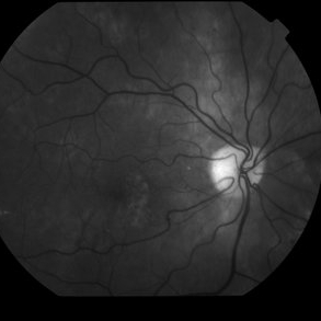
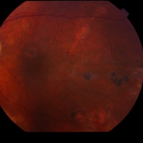 West Nile Virus Choroiditis
Apr 4 2014 by Suber S. Huang, MD, MBA, FASRS
Fundus photograph (11 month follow-up) of an 88-year-old woman who developed West Nile virus encephalitis on 08/2012 and subsequent choroiditis.
Photographer: Geoffrey Pankhurst; University Hospitals Eye Institute, Case Western Reserve University, Cleveland, OH
Imaging device: TopCon TRC50EX
Condition/keywords: choroiditis, disseminated choroiditis, infectious uveitis, optic nerve atrophy
-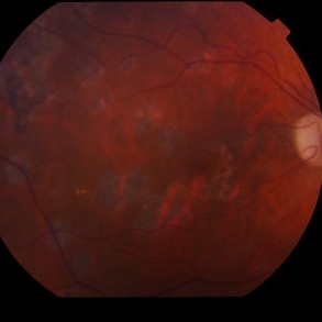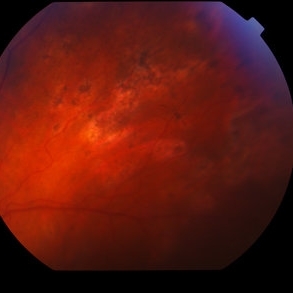
 West Nile virus choroiditis
Apr 4 2014 by Suber S. Huang, MD, MBA, FASRS
Fundus photograph (11 month follow-up) of an 88-year-old woman who developed West Nile virus encephalitis on 08/2012 and subsequent choroiditis
Photographer: Geoffrey Pankhurst; University Hospitals Eye Institute, Case Western Reserve University, Cleveland, OH
Imaging device: TopCon TRC50EX
Condition/keywords: choroiditis, disseminated choroiditis, infectious uveitis, optic nerve atrophy, West Nile virus choroiditis
-
 West Nile Virus Choroiditis
Apr 4 2014 by Suber S. Huang, MD, MBA, FASRS
Fundus photograph (11 month follow-up) of an 88-year-old woman who developed West Nile virus encephalitis on 08/2012 and subsequent choroiditis.
Photographer: Geoffrey Pankhurst; University Hospitals Eye Institute, Case Western Reserve University, Cleveland, OH
Imaging device: TopCon TRC50EX
Condition/keywords: choroiditis, disseminated choroiditis, infectious uveitis, optic nerve atrophy
 Loading…