Search results (31 results)
-
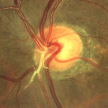
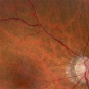
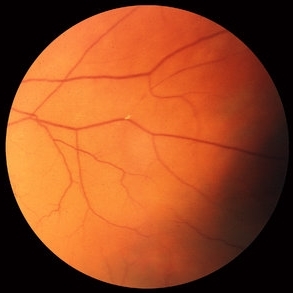
 A Glow in the Darkness : Hollenhorst Plaque
Aug 21 2023 by Harsh Vardhan Singh, MS
82-year-old-female with a history of some disturbance of vision in the left eye with the finding of hollenhorst plaque in one of the branches of central retinal artery
Photographer: Dr Harsh Vardhan Singh
Condition/keywords: hollenhorst plaque
-
 A Glow in the Darkness: Hollenhorst Plaque
Aug 22 2023 by Harsh Vardhan Singh, MS
82 year-old-female with a history of some disturbance of vision in the left eye with the finding of hollenhorst plaque in one of the branches of central retinal artery
Photographer: Dr Harsh Vardhan Singh
Condition/keywords: hollenhorst plaque
-
 Brach Retinal Artery Occlusion
Oct 2 2013 by Jerald A. Bovino, MD
There is a hollenhorst plaque causing a branch retinal artery occlusion. The patient has scars from prior panretinal laser photocoagulation.
Condition/keywords: branch retinal artery occlusion (BRAO), hollenhorst plaque, pan-retinal photocoagulation (PRP)
-
 Branch Retinal Artery Occlusion
Mar 27 2018 by Nichole Lewis
Branch retinal artery occlusion with a Hollenhorst Plaque.
Photographer: Nichole Lewis
Condition/keywords: branch retinal artery occlusion (BRAO), hollenhorst plaque
-
 Branch Retinal Artery Occlusion
Mar 27 2018 by Nichole Lewis
Branch retinal artery occlusion with a Hollenhorst Plaque.
Photographer: Nichole Lewis
Condition/keywords: branch retinal artery occlusion (BRAO), hollenhorst plaque
-
 Branch Retinal Artery Occlusion
Oct 2 2013 by Jerald A. Bovino, MD
There is a hollenhorst plaque causing a branch retinal artery occlusion. The patient has scars from prior panretinal laser photocoagulation.
Condition/keywords: branch retinal artery occlusion (BRAO), hollenhorst plaque, pan-retinal photocoagulation (PRP)
-
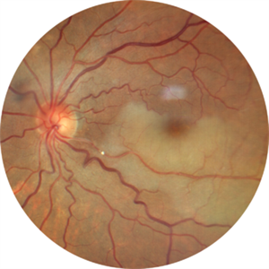
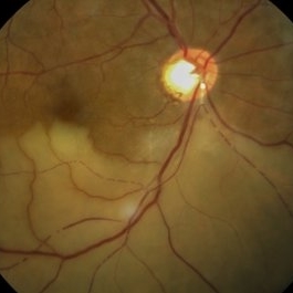 BRAO with Hollenhorst plaque
Jul 22 2021 by Vishal Gupta, MBBS, MS
Fundus image of 54-year-old male patient with inferior branch retinal artery occlusion and a prominent Hollenhorst plaque seen as shining white dot at disk along with cattle trucking phenomenon.
Photographer: Dr Vishal Gupta, INHS Asvini, Mumbai, INDIA
Imaging device: Zeiss
Condition/keywords: branch retinal artery occlusion (BRAO), hollenhorst plaque
-
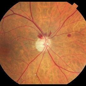
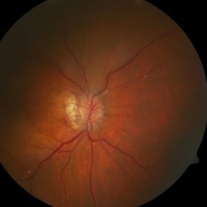
 Central Retinal Artery Occlusion (CRAO)
Dec 27 2016 by Manish Nagpal, MD, FRCS (UK), FASRS
Acute CRAO with hollenhorst plaque.
Photographer: hardik Jain
Condition/keywords: central retinal artery occlusion (CRAO), edema, hollenhorst plaque, retinal infarction
-
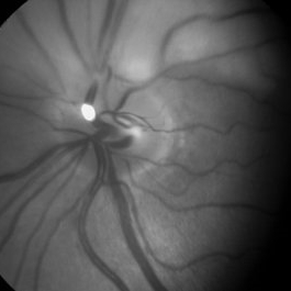
 HHPlaqueON
Aug 13 2021 by Jeffrey Barker
Hollenhorst Plaque
Photographer: Jeffrey P. Barker, B.S. Retina Vitreous Surgeons of C.N.Y.
Condition/keywords: hollenhorst plaque, optic nerve
-
 Hollenhorst Plaque
Jun 11 2023 by Ethan K Sobol, MD
Hollenhorst plaque located at an arterial bifurcation along the inferior arcade
Condition/keywords: atherosclerosis, embolus, hollenhorst plaque
-
 Hollenhorst Plaque
Jul 25 2021 by Vishal Gupta, MBBS, MS
Hollenhorst plaque shining in the red free image of a patient with superior branch retinal artery occlusion.
Photographer: Dr Vishal Gupta, INHS Asvini, Mumbai, INDIA
Imaging device: Zeiss
Condition/keywords: hollenhorst plaque, red-free
-
 Hollenhorst Plaque
Jun 25 2024 by Virginia Gebhart
75 year female with complaint of shadow in the bottom of her vision for many years. Hollenhorst plaque on superior pole of the disc and sclerotic superotemporal arteriole. Also DBHs superiorly most likely due to combined BRAO/BRVO.
Photographer: Virginia Gebhart
Imaging device: Topcon 50DX
Condition/keywords: branch retinal artery occlusion (BRAO), branch retinal vein occlusion (BRVO), hollenhorst plaque, sclerotic arteriole
-
 Hollenhorst Plaque
Sep 21 2023 by Ben Serar
Fundus photograph showing multiple whitish intra-arterial dot like deposits suggestive of Hollenhorst plaques.
Condition/keywords: hollenhorst plaque
-
 Hollenhorst Plaque
Sep 2 2025 by KANWALJEET HARJOT MADAN, M.S. (Ophthalmology); FAICO (Vitreous - Retina)
A 64 year-old male presented with sudden decrease in vision in LE for 1 week. His BCVA in LE was 20/200. Fundus exam revealed presence of whitish ischemic area in macula superior to fovea suggestive of branch retinal artery occlusion. A bright tiny refractile cholesterol embolus (Hollenhorst plaque) was visible in retinal artery. The patient was advised cardiology consultation.
Photographer: Dr. Kanwaljeet Harjot Madan, Thind Eye Hospital, Jalandhar City (Punjab). INDIA.
Imaging device: Zeiss Fundus Camera
Condition/keywords: branch retinal artery occlusion (BRAO), hollenhorst plaque
-
---thumb.jpg/image-square;max$300,300.ImageHandler) Hollenhorst Plaque
Hollenhorst Plaque
-
---thumb.jpg/image-square;max$300,300.ImageHandler) Hollenhorst Plaque
Hollenhorst Plaque
-
 Hollenhorst Plaque
Oct 8 2012 by David R. Chow, MD, FRCS(C)
Condition/keywords: hollenhorst plaque
-
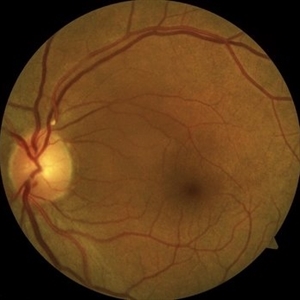
 Hollenhorst Plaque
Sep 18 2016 by John T. Thompson, MD
Color photo of Hollenhorst plaque at branch of inferotemporal artery.
Imaging device: Zeiss FF4
Condition/keywords: branch retinal artery occlusion (BRAO), hollenhorst plaque
-
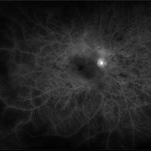
 Hollenhorst Plaque
Sep 18 2016 by John T. Thompson, MD
Fluorescein angiogram showing branch retinal artery occlusion at branch of inferotemporal artery.
Imaging device: Zeiss FF4
Condition/keywords: branch retinal artery occlusion (BRAO), hollenhorst plaque
-
 Hollenhorst Plaque in Eye with BRAO
Oct 1 2012 by Jeffrey G. Gross, MD, FASRS
Hollenhorst plaque in eye with BRAO.
Condition/keywords: branch retinal artery occlusion (BRAO), hollenhorst plaque
-
 Hollenhorst Plaque in Eye with CRAO
Oct 1 2012 by Jeffrey G. Gross, MD, FASRS
Hollenhorst plaque in eye with CRAO.
Condition/keywords: central retinal artery occlusion (CRAO), hollenhorst plaque, in eye
-
 Hollenhorst Plaques, multiple
Feb 20 2013 by From the Collections of Thomas M. Aaberg, MD and Thomas M. Aaberg Jr., MD
No history.
Condition/keywords: hollenhorst plaque
-
 HOLLENHORST-OE
-
 Ocular Ischemic Syndrome
Jun 18 2025 by Korey Starkey
58-year-old patient with OIS and Hollenhorst plaque.
Photographer: Korey Starkey
Imaging device: Optos
Condition/keywords: capillary nonperfusion, fluorescein angiogram (FA), hollenhorst plaque, NVD, ocular ischemic syndrome, Optos
-
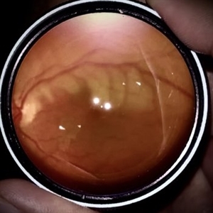 Branch Retinal Artery Occlusion
Oct 1 2024 by Angel Enrique Flores Pineda
Fundus photograph of a 78-year-old woman with poorly controlled systemic arterial hypertension and dyslipidemia. Hollenhorst plaque can be observed.
Photographer: Angel Enrique Flores Pineda, Hospital General de Zona #20
Imaging device: Smartphone (IPhone 15 plus)
Condition/keywords: branch retinal artery occlusion (BRAO)
 Loading…