Search results (131 results)
-
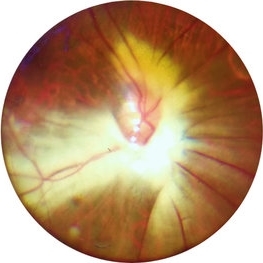
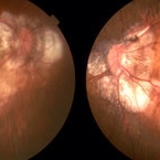
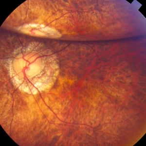 "Internal Mirroring" Effect by Intraocular Gas
Mar 25 2014 by Homayoun Tabandeh, MD, FASRS
"Internal mirroring" by residual intraocular gas in a highly myopic patient 3 weeks post repair of retinal detachment with pars plana vitrectomy and C3F8 gas.
Photographer: Danny Rivas
Condition/keywords: high myopia, intraocular gas
-
 Bilateral CNV in High Myopia
Apr 2 2019 by Gary R. Cook, MD, FACS
Right eye of a 60-year-old white female with -9D myopia, myopic maculopathy, and visible (Type 1) CNV; V.A. = 20/40.
Imaging device: Topcon VT-50
Condition/keywords: choroidal neovascular membrane (CNVM), choroidal neovascularization (CNV), high myopia, myopic degeneration, myopic fundus, pathologic myopia
-
 Bilateral CNV in High Myopia
Apr 2 2019 by Gary R. Cook, MD, FACS
Left eye of a 60-year-old white female with -9D myopia and bilateral visible (Type 1) CNV; V.A. = 20/30.
Imaging device: Topcon VT-50
Condition/keywords: choroidal neovascular membrane (CNVM), choroidal neovascularization (CNV), high myopia, myopic degeneration, myopic fundus, pathologic myopia
-
 Buckled Eye in a case of High Myopia
Jan 8 2020 by Sham Talati, DOMS
Buckled Eye in a case of high myopia.
Photographer: Dr. Sham Talati,Retina Foundation,Ahmedabad
Imaging device: Nidek Mirante
Condition/keywords: high myopia, scleral buckle
-
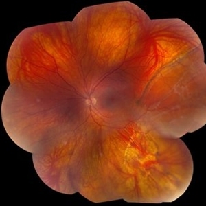 Chronic Retinal Detachment in a Young Myopic Patient
Nov 6 2019 by Kamal Kishore, MD, MBBS
Chronic retinal detachment in a 27-year-old myopic female showing spontaneous reattachment in inferotemporal quadrant, and demarcation line and subretinal gliosis in superotemporal quadrant.
Photographer: Stephanie Shaver
Imaging device: Topcon 50 EX with OIS Winstation
Condition/keywords: chronic retinal detachment, high myopia
-
 CNVM due to Pathologic Myopia
Apr 2 2019 by Gary R. Cook, MD, FACS
55-year-old Asian male with -9.50D myopia with a visible (Type I) CNVM and thin hemorrhage in the macula: V.A.= 20/200
Imaging device: Topcon VT-50
Condition/keywords: choroidal neovascular membrane (CNVM), high myopia, pathologic myopia, retinal hemorrhage
-
 CNVM Due to Pathologic Myopia
Apr 2 2019 by Gary R. Cook, MD, FACS
Fluorescein angiogram frame of the left eye of the 55-year-old Asian male with -9.50D myopia OS with a subfoveal CNVM and hemorrhage secondary to high myopia; V.A.= 20/200.
Imaging device: Topcon VT-50
Condition/keywords: choroidal neovascular membrane (CNVM), high myopia, pathologic myopia, retinal hemorrhage, subfoveal choroidal neovascularization
-
---thumb.jpg/image-square;max$300,300.ImageHandler) Congenital Cataracts
Congenital Cataracts
Dec 26 2013 by David Callanan, MD
42-year-old female patient, 20/30 OU; high myope.
Condition/keywords: congenital cataract, high myopia
-
---thumb.jpg/image-square;max$300,300.ImageHandler) Congenital Cataracts
Congenital Cataracts
Dec 26 2013 by David Callanan, MD
42-year-old female patient, 20/30 OU; high myope.
Condition/keywords: congenital cataract, high myopia
-
---thumb.jpg/image-square;max$300,300.ImageHandler) Congenital Cataracts
Congenital Cataracts
Dec 26 2013 by David Callanan, MD
42-year-old female patient, 20/30 OU; high myope.
Condition/keywords: congenital cataract, high myopia
-
---thumb.jpg/image-square;max$300,300.ImageHandler) Congenital Cataracts
Congenital Cataracts
Dec 26 2013 by David Callanan, MD
42-year-old female patient, 20/30 OU; high myope.
Condition/keywords: congenital cataract, high myopia
-
---thumb.jpg/image-square;max$300,300.ImageHandler) Congenital Cataracts
Congenital Cataracts
Dec 26 2013 by David Callanan, MD
42-year-old female patient, 20/30 OU; high myope.
Condition/keywords: congenital cataract, high myopia
-
---thumb.jpg/image-square;max$300,300.ImageHandler) Congenital Cataracts
Congenital Cataracts
Dec 26 2013 by David Callanan, MD
42-year-old female patient, 20/30 OU; high myope.
Condition/keywords: congenital cataract, high myopia
-
---thumb.jpg/image-square;max$300,300.ImageHandler) Congenital Cataracts
Congenital Cataracts
Dec 26 2013 by David Callanan, MD
42-year-old female patient, 20/30 OU; high myope.
Condition/keywords: congenital cataract, high myopia
-
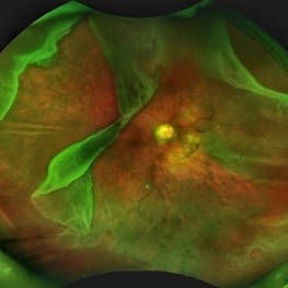
 Degenerative Myopia
Apr 12 2023 by Ahmed Abbas Hashmi, OD
Right eye Fundus photograph of a 61-year-old female with pathological myopia.
Condition/keywords: chorioretinal atrophy, high myopia, pathologic myopia
-
 Dome Shaped Macula
Jan 7 2025 by Jordyn Beckman
Autofluorescence photograph of 36 year old woman with Dome Shaped Macula with hypoautofluorescence and hyperautofluorescence centrally.
Photographer: Jordyn Beckman
Imaging device: California Optos
Condition/keywords: autoflorescence, convex protrusion, dome shaped macula, high myopia, hyperautofluorescent centrally, hypoautofluorescence
-
 Dome-Shaped Macula With Subretinal Fluid
Jun 14 2018 by Gerardo Garcia-Aguirre, MD
EDI OCT of the right eye of a 17-year-old highly myopic girl. Subfoveal fluid is present. There is choroidal thinning, and scleral thickening in the foveal area.
Photographer: Gerardo Garcia-Aguirre, MD
Imaging device: Heidelberg Spectralis
Condition/keywords: dome shaped macula, high myopia
-
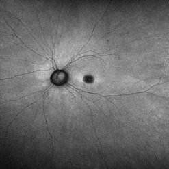
 Extensive Myelinated Nerve Fibres
May 20 2021 by Anmol Naik
A 21-year-old Indian male presented with incidentally discovered subnormal vision in the left eye. On examination, he had esotropia with high myopia of -14 dioptres. Fundus examination revealed extensively myelinated nerve fibres around the optic disc extending along the arcade but sparing the fovea. The association of myelinated nerve fibres with high myopia and amblyopia is well documented but the causal association between these is unproven. Early detection of refractive error and aggressive therapy to prevent amblyopia has been reported with some success.
Photographer: Anmol Naik, Nakshatra Superspeciality Eye Hospital, Pune, India.
Imaging device: Zeine slit-lamp mounted Fundus imaging system
Condition/keywords: amblyopia, high myopia, myelinated nerve fibers
-
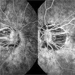 Fluorescein Angiography in High Myopia
Dec 7 2019 by Anfisa Ayalon, MD
Fluorescein angiography pictures of a 55-year-old woman with high myopia.
Photographer: Anfisa Ayalon, MD., Meir Medical Center, Kfar Saba, Israel.
Condition/keywords: fluorescein angiogram (FA), high myopia, peripapillary atrophy
-
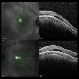 Foveal Detachment in Dome-Shaped Macula
May 6 2017 by Mitzy E Torres Soriano, MD
Vertical scans of optical coherence tomography showing a foveal detachment at the top of dome-shaped macula in a 48-year-old female with high myopia.
Photographer: Mitzy E. Torres Soriano
Condition/keywords: detachment, high myopia, macula
-
 Foveoschisis secondary to high myopia
Mar 13 2015 by Niloofar Piri, MD
Infrared and HD-OCT of the right eye in a 55-year-old African American female with high myopia (more than -6.00 D), BCVA: 20/25 OU Cartwheel appearance of the fovea in the infrared imaging is visible. HD- OCT demonstartes schisis in different layers of the retina (both NFL and OPL; notice stretching of the Muller cells); VMT is also present . Outer retinal layers are preserved which explains the good vision . She had the same findings in OS.
Photographer: Niloofar Piri, MD
Imaging device: Heidelberg Spectralis
Condition/keywords: high myopia, retinoschisis
-
 Fuch's Spot
Apr 2 2019 by Gary R. Cook, MD, FACS
20-year-old patient with high myopia and a Fuch's spot OD.
Condition/keywords: Fuchs, high myopia, pathologic myopia
-
 Giant Retinal Tear
Oct 9 2012 by Audina M. Berrocal, MD FASRS
Teenager with high myopia and a GRT
Photographer: Ditte Hess CRA, BPEI
Imaging device: Fundus Camera
Condition/keywords: high myopia, retinal degeneration, retinal tear
-
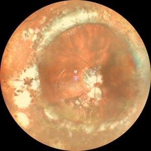
 Giant Retinal Tear with Multiple Retinal Breaks
Apr 21 2025 by Hrishikesh Naik, MS
A 28 year old high myope with retinal detachment associated with a supero-temporal giant retinal tear in addition to multiple peripheral horseshoe tears and an additional supero-nasal retinal tear.
Photographer: Hrishikesh Naik
Imaging device: Optos Daytona
Condition/keywords: giant retinal tear, High Myopia, horseshoe tear, retinal break, retinal detachment
-
 High Myopia
Sep 24 2020 by Anthony Maida
59-year-old female with high myopia.
Photographer: Anthony C. Maida, Hospital Central Militar, Ciudad de México
Imaging device: Topcon TRC-NW8
Condition/keywords: high myopia
 Loading…