Search results (91 results)
-
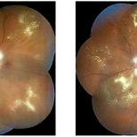
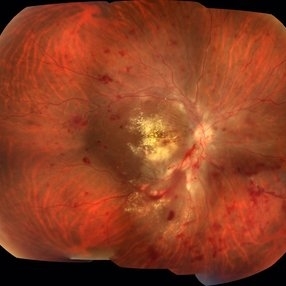
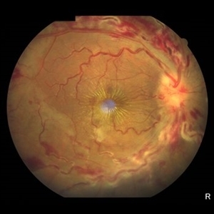 Central Retinal Vein Occlusion
Jun 21 2025 by Moazzam Parvez
Fundus photograph of a 56 year old male presenting with dilated tortuous vessels with adjoining Hard exudates and macular star.
Photographer: Moazzam Parvez , Netralayam , Kolkata
Imaging device: Topcon Maestro 2
Condition/keywords: CRVO with macular edema, hard exudates, macular star
-
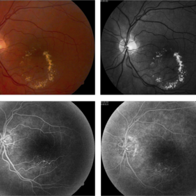
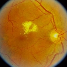 Hard exudates
Sep 21 2023 by Ben Serar
Fundus photograph of RE showing hard exudates at the macula, superior to the fovea.
Condition/keywords: hard exudates
-
 Hypertensive Retinopathy
Sep 12 2023 by Ben Serar
Fundus photograph of LE showing Disc edema with optic disc pallor, hard exudates with dot-blot haemorrhages at the macula ,along with arteriolar attenuation, in a case of Hypertensive retinopathy.
Condition/keywords: arteriolar attenuation, disc edema, Hard exudates, hypertensive retinopathy
-
 Leaking Aneurysms in Diabetic Retinopathy
Mar 22 2024 by Vaidehi Sathaye
Fundus photograph of LE of a 50 year old female with leaking aneurysms encircled by hard exudates, as a sequelae of Diabetic Retinopathy.
Photographer: Dr. Vaidehi Sathaye
Imaging device: Topcon
Condition/keywords: aneurysm, diabetic retinopathy, hard exudates
-
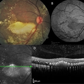 Macular Edema
Jun 5 2021 by Aditya Verma, MBBS, MS, Post fellow
Multimodal imaging of a 36-year-old male with peripheral vasoproliferative tumor. Posterior pole showed exquisite pattern of hard exudate accumulation in petaloid pattern (A), fundus autofluorescence (B) and infrared image (C) portrayed the precise pattern of hard exudate distribution; Optical coherence tomography scan (D) showed a uniformly distributed parallel clumps of exudates in the outer plexiform layer.
Photographer: Aditya Verma
Imaging device: Heidelberg Spectralis
Condition/keywords: hard exudates, macular edema, macular exudates
-
 NPDR With Myelinated Nerve Fibers
Nov 5 2018 by Diva Kant Misra, MBBS, DO, DNB, MNAMS, FVRS
Bilateral montage funds photo images of a 56-year-old diabetic patient showing signs of NPDR along with myelinated nerve fibers.
Photographer: Hiteshwar Saikia
Condition/keywords: diabetes, hard exudates, myelinated nerve fibers, nonproliferative diabetic retinopathy
-
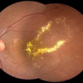
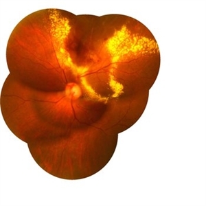 RAM With Garland of Hard Exudate
Mar 3 2020 by KRISHNENDU NANDI, MS
Fundus Photo of left eye of 75-year-old female with retinal artery macroaneurysm at superior quadrant with garland like hard exudates.
Photographer: KRISHNENDU NANDI
Imaging device: Topcon
Condition/keywords: hard exudates, retinal arterial macroaneurysm
-
 RAMA
RAMA
Sep 7 2018 by John S. King, MD
80 yo WF with a history of HTN, CAD, and brain aneurysm referred for possible BRVO OS and one month decreased vision. 20/20 OD and 20/100 OS. Six months since light laser applied to super-temporal MA. Retinal exudates and juxtafoveal exudative scar seen here (OCT showed resolution of CME and SRF)
Photographer: Stacey Coleman
Imaging device: Topcon
Condition/keywords: foveal hard exudates, hard exudates, ruptured macroaneurysm
-
 Vascular Anormalities
Jan 6 2016 by Andrea Arriola-Lopez, MD MSc
77-year-old man. Decrease of visual acuity OS. VA 20/30 IOP 14mmHg. Fundus examination findings: Hard exudates, microaneurysms near to fovea. OCT shows IRF. Late leakage on FA.
Photographer: Andrea Elizabeth Arriola-Lopez, MSc MD
Condition/keywords: abnormal retinal vessel, aneurysm, hard exudates, vascular anomaly
-
---thumb.JPG/image-square;max$300,300.ImageHandler) Exudative Diabetic Maculopathy
Exudative Diabetic Maculopathy
Nov 18 2013 by Mallika Goyal, MD
Hard exudates at the macula, including the foveal centre, in an eye with diabetic retinopathy.
Photographer: Mallika Goyal, MD, Apollo Health City, Hyderabad
Condition/keywords: exudative diabetic retinopathy
-
---thumb.JPG/image-square;max$300,300.ImageHandler) Exudative Diabetic Maculopathy
Exudative Diabetic Maculopathy
Nov 18 2013 by Mallika Goyal, MD
Hard exudates at macula, including at foveal centre, in an eye with diabetic retinopathy.
Photographer: Mallika Goyal, MD, Apollo Health City, Hyderabad
Condition/keywords: exudative diabetic retinopathy
-
---thumb.JPG/image-square;max$300,300.ImageHandler) Exudative Diabetic Maculopathy
Exudative Diabetic Maculopathy
Nov 18 2013 by Mallika Goyal, MD
Hard exudates at the fovea in an eye with NPDR.
Photographer: Mallika Goyal, MD, Apollo Health City, Hyderabad
Condition/keywords: exudative diabetic retinopathy
-
 A Fleet of Boat-Shaped Hemorrhages
Aug 1 2024 by James P Dossett, MD
Pseudocolor fundus photograph of the left eye of a 54-year-old diabetic man presenting with bilateral vision loss. Examination revealed 20/200 vision OS with extensive preretinal and vitreous hemorrhage, marked diffuse neovascularization, macular edema and hard exudates.
Photographer: Beth Smith, West Virginia University Eye Institute
Condition/keywords: proliferative diabetic retinopathy (PDR)
-
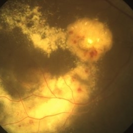 Abundant Hard Exudates - Diabetic Macular Edema
Oct 3 2013 by Gerardo Garcia-Aguirre, MD
Abundant hard exudates - diabetic macular edema.
Condition/keywords: diabetic macular edema
-
 Acute Necrotizing Retinal Vasculitis as Onset of Systemic Lupus Erythematosus.
Sep 3 2016 by ADRIANO FERREIRA
A 28-year-old white man was referred to the rheumatology clinic with gradually and rapid deterioration of the vision (both eyes). In this picture, we can observe cotton wool spots in the papillomacular area and extensive hemorrhages in posterior polo and in the middle periphery. Hard exudates are present in macular area (macular edema)
Photographer: Claudio Zett Lobo
Imaging device: TRC50DXi TOPCON
Condition/keywords: systemic lupus erythematosus (SLE) vasculitis, vasculitis
-
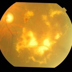
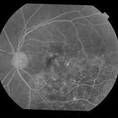
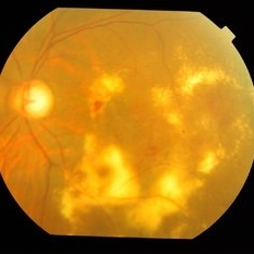 Adult Coats' Disease
Aug 18 2015 by Mallika Goyal, MD
Left fundus of a 61-year-old non diabetic, non hypertensive lady complaining of vision deterioration for 1 year showing massive hard exudates at the macula. Fluorescein angiography revealed microvascular abnormalities over the posterior pole and temporal midperiphery and extensive capillary non-perfusion over the temporal retinal quadrants. OCT revealed macular edema. Fellow eye fundus and angiogram were normal.
Photographer: Mallika Goyal, MD, Apollo Health City, Jubilee Hills, Hyderabad
Condition/keywords: Coats' disease
-
 Adult Coats' Disease
Aug 18 2015 by Mallika Goyal, MD
Left fundus of a 61-year-old non diabetic, non hypertensive lady complaining of vision deterioration for 1 year showed massive hard exudates at the macula. Fluorescein angiography revealed microvascular abnormalities over the posterior pole and temporal midperiphery and extensive capillary non-perfusion over the temporal retinal quadrants. OCT revealed macular edema. Fellow eye fundus and angiogram were normal.
Photographer: Mallika Goyal, MD, Apollo Health City, Jubilee Hills, Hyderabad
Condition/keywords: Coats' disease
 Loading…