Search results (179 results)
-
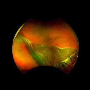
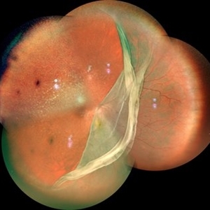
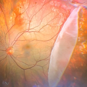
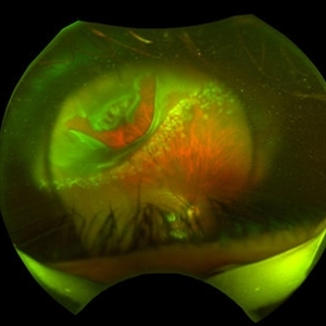 "Hang in There"
Apr 20 2021 by Tomas Minelli, MD
Fundus wide field photograph of a 50-year-old man with a macular detachment associated with a big temporal superior tear. The laser is firmly holding the progression of the tear in the 14th day post- laser. BCVA 20/20
Photographer: Livia Conci, Universtity of São Paulo
Imaging device: Optos Daytona
Condition/keywords: giant retinal tear
-
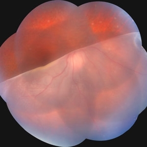
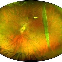
 24 Hours Post Scleral Wound Closure+ Scleral Buckle+25 g Vitrectomy+Silicon Oil
Jan 23 2015 by Carlos Quezada-Ruiz, MD, FASRS
24 hours post op fundus photograph of a 43-year-old man who had perforating injury to the right eye with a small piece of plastic while he was hammering. OD LP, subconjunctival hemorrhage, clear cornea, hyphema, irido and ciclodyalisis as well as a luxated lens with traumatic cataract and a dense vitreous hemorrhage. B-US showed rhegmatogenous retinal detachment with a tear and a big inferior hemorrhagic choroidal detachment. 360 peritomy revealed 2-entry scleral wounds were found in zone II (M V and M VI) and closure was performed. 25 G PPV was performed with the infusion canal placed in the AC through the limbus. Lensectomy and removal of a dense recent vitreous hemorrhage revealed a white detached retina with an exit wound through the temporal inferior segment of the optic nerve with a nasal GRT and sub retinal hemorrhage as well as temporal inferior choroidal, PVD was induced and PFOs helped stabilizing the retina while vitrectomy and sub-retinal hemorrhage was removed through the GRT. Fluid air exchange was made and 360 endolaser over the buckle indentation was done and silicon oil was used as endotamponade. This picture was taken 24 hrs after the surgery.
Photographer: Lilibeth Rodriguez, Instituto de la Visión. Torreon, Mexico.
Condition/keywords: central retinal artery occlusion (CRAO), giant retinal tear, trauma
-
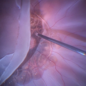
Flattening of Giant retinal tear
Oct 24 2022 by Manish Nagpal, MD, FRCS (UK), FASRS
This video highlights the step of eversion of the giant retinal tear flap and flattening of retina using heavy PFCL liquid
Photographer: Manish Nagpal
Condition/keywords: flap, giant retinal tear, GRT, PFCL, video, vitrectomy
-
 Flattening Out the Retina With PFCL
Sep 28 2024 by Anjana Mirajkar, MS Ophthalmology
An intra operative image showing slow injection of PFCL to flatten out the folded retina.
Photographer: Dr. Anjana Mirajkar -Retina Foundation, Ahmedabad
Condition/keywords: GIANT RETINAL TEAR, PFCL
-
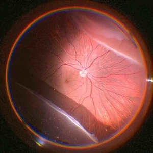
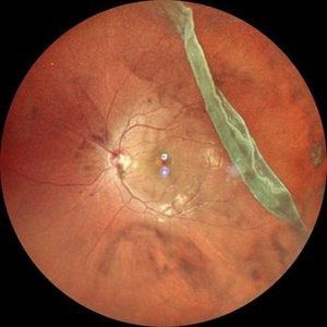 Giant Retinal Tear
Oct 11 2024 by Anjana Mirajkar, MS Ophthalmology
Widefield fundus photograph of LE showing giant retinal tear extending from 12 to 4 o clock.
Photographer: Dr. Anjana Mirajkar -Retina Foundation, Ahmedabad
Imaging device: Mirante-Nidek
Condition/keywords: giant retinal tear
-
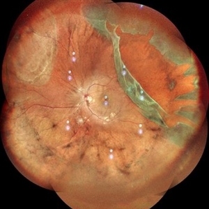 Giant Retinal Tear
Oct 11 2024 by Anjana Mirajkar, MS Ophthalmology
Fundus photograph montage of LE showing a giant retinal extending from 12 to 4 o clock.
Photographer: Dr. Anjana Mirajkar -Retina Foundation, Ahmedabad
Imaging device: Mirante-Nidek
Condition/keywords: GIANT RETINAL TEAR
-
 Giant Retinal Tear
May 20 2024 by Aysha AlOqab, MB BCh BAO
Fundus photograph of a 40-year-old man who presented with a history of progressive inferior visual field defect in the right eye over 2-3 weeks.
Photographer: Saleh AlDhafiri, King Khaled Eye Specialist Hospital, Riyadh, KSA
Imaging device: Optos
Condition/keywords: giant retinal tear, idiopathic
-
 Giant Retinal Tear
Jul 15 2024 by Arthi Mohankumar , MS,MRCS ED, FICO,FAICO
Fundus montage of a 15 year old boy with Marfans syndrome who presented with defective vision in the right eye.
Photographer: Arthi Mohankumar
Condition/keywords: giant retinal tear, Retinal detachment
-
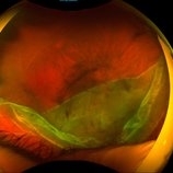
 Giant Retinal Tear
Mar 15 2022 by Jesus Lozano, MD
42 year old woman with a supero temporal giant retinal tear.
Photographer: Dr. Mohamad Midlij
Imaging device: Optos Silverstone
Condition/keywords: retinal detachment with retinal defect, retinal tear
-
 Giant retinal Tear
Apr 26 2022 by Jeffrey Barker
Giant retinal Tear
Photographer: Jeffrey P. Barker B.S.
Condition/keywords: retinal tear
-
 Giant Retinal Tear
Dec 26 2022 by Vaidehi Sathaye
Fundus photograph of RE of a 61 year male patient with a Giant Retinal Tear
Photographer: Dr. Vaidehi Sathaye
Imaging device: Mirante
Condition/keywords: giant retinal tear
-
 Giant Retinal Tear
Apr 21 2022 by Vaidehi Sathaye
Fundus photograph of a 16 year old male with a Rhegmatogenous Retinal Detachment secondary to a Giant Retinal Tear in the right eye.
Photographer: Dr. Vaidehi Sathaye, Retina Foundation
Condition/keywords: giant retinal tear
-
 Giant Retinal Tear
Apr 19 2022 by Thais Bastos
A 44-year-old female patient with sudden loss of visual acuity in her left eye. Note retinal detachment with giant retinal tear with the retina folded over itself.
Photographer: Thaís Azeredo Bastos - HCRP-USP, Brazil
Imaging device: Optos California
Condition/keywords: giant retinal tear
-
 Giant Retinal Tear
Oct 14 2022 by Angela Rico
40 year old female who presented with Giant Retinal tear
Photographer: Angela Rico M.D.
Imaging device: OCT
Condition/keywords: retinal tear
-
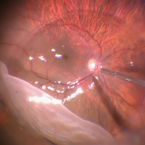
 Giant Retinal Tear
Sep 28 2024 by Anjana Mirajkar, MS Ophthalmology
An intra operative image of the right eye showing a giant retinal tear with a superior retinal detachment.
Photographer: Dr. Anjana Mirajkar -Retina Foundation, Ahmedabad
Condition/keywords: GIANT RETINAL TEAR
-
 Giant Retinal Tear
Sep 28 2024 by Anjana Mirajkar, MS Ophthalmology
An intra-operative still showing giant retinal tear from 6 to 11 clock hour with folded posterior margins.
Photographer: Dr. Anjana Mirajkar -Retina Foundation, Ahmedabad
Condition/keywords: GIANT RETINAL TEAR
-
 Giant Retinal Tear
Sep 28 2024 by Anjana Mirajkar, MS Ophthalmology
An intra operative image showing slow injection of PFCL on the posterior pole to unfold the posterior margins in case of giant retinal tear.
Photographer: Dr. Anjana Mirajkar -Retina Foundation, Ahmedabad
Condition/keywords: GIANT RETINAL TEAR
-
 Giant Retinal Tear
Feb 20 2024 by Soobien Lee
Optos color fundus photograph of a 40-year-old caucasian male who is a UFC fighter with a total retinal detachment in his right eye secondary to a giant retinal tear from 10 o'clock to 2 o'clock.
Photographer: Trinity Wolf, Elman Retina Group
Imaging device: Optos Ultra-Widefield Imaging
Condition/keywords: giant retinal tear, optos, Retinal Detachment, Retinal tear with detachment, trauma
-
 Giant Retinal Tear
May 15 2014 by Manish Nagpal, MD, FRCS (UK), FASRS
Patient presenting with a acute loss of vision with a giant retinal tear.
Photographer: pooja barot, Optometrist, Retina Foundation, Ahmedabad
Condition/keywords: giant retinal tear
-
 Giant Retinal Tear
Jan 11 2022 by Manish Nagpal, MD, FRCS (UK), FASRS
Intraoperative photo a temporal giant retinal tear with everted flap and some laser marks noted on the bare choroid from previous barrage attempt elsewhere.
Photographer: Manish Nagpal, Retina Foundation, Ahmedabad, India
Imaging device: Sony PMW -10 MD surgical camera
Condition/keywords: giant retinal tear
-
 Giant retinal tear
Nov 6 2019 by Veronica A. Kon Graversen, MD
"Taquito dorado retina." Photograph of a 49-year-old Hispanic male with macula-on retinal detachment secondary to a giant retinal tear.
Photographer: Alex Romera
Imaging device: Optos
Condition/keywords: giant retinal tear
-
 Giant Retinal Tear
May 27 2020 by Jamin S. Brown, MD
Fundus photo montage of 55-year-old male with retinal detainment and giant retinal tear. Same patient as above.
Photographer: Stefanie Palmer CRA, Retina-Vitreous Surgeons of CNY
Condition/keywords: giant retinal tear
-
 Giant Retinal Tear
May 27 2020 by Jamin S. Brown, MD
Fundus photo montage of 55-year-old male with retinal detachment and giant retinal tear.
Photographer: Stefanie Palmer CRA, Retina-Vitreous Surgeons of CNY
Condition/keywords: giant retinal tear
-
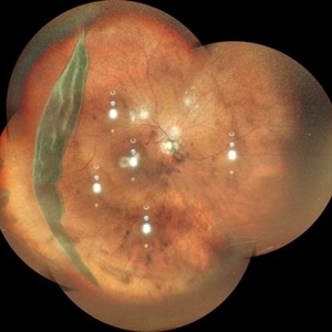
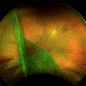
 Giant Retinal Tear
Mar 29 2014 by Min Kim, MD, PhD, MBA, FASRS
Wide field fundus photograph of a 25 year-old male shows giant retinal tear with inverted retinal flap.
Photographer: Young Duk Bae, Yonsei University, Gangnam Severance Hospital
Imaging device: Optomap
Condition/keywords: giant retinal tear
-
---thumb.JPG/image-square;max$300,300.ImageHandler) Giant Retinal Tear
Giant Retinal Tear
Jul 13 2013 by Jason S. Calhoun
Giant retinal tear with retinal detachment.
Photographer: Jason S. Calhoun, Department of Ophthalmology, Mayo Clinic Jacksonville, Florida
Condition/keywords: retinal tear
 Loading…