Search results (9 results)
-
 Choroidal Infarcts Post Preeclampsia
Aug 26 2012 by Andrew N. Antoszyk, MD FASRS
Condition/keywords: choroidal infarction, preeclampsia
-
 Choroidal Infarcts Preeclampsia
Aug 26 2012 by Andrew N. Antoszyk, MD FASRS
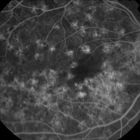
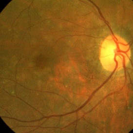
66-year-old Hispanic female with a history of severe preeclampsia at age 45. This photograph shows faint gray choroidal scars. Seen better on FA (in gallery).
Condition/keywords: choroidal infarction, preeclampsia
-
 Hypertensive Retinopathy
May 1 2024 by Marco Antonio Sauza
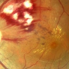
36 year old male with uncontrolled systemic hypertension.
Photographer: MARCO SAUZA CASTILLEJOS
Imaging device: VISUCAM ZEISS
Condition/keywords: choroidal infarction, hypertensive retinopathy, macular star
-
 Hypertensive Retinopathy Grade IV
May 1 2024 by Marco Antonio Sauza
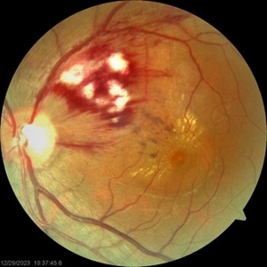
Fundus photograph of an 36-year-old male with uncontrolled systemic hypertension, >200/100mmhg, presenting decreased vision in the left eye.
Photographer: MARCO SAUZA CASTILLEJOS
Imaging device: VISUCAM ZEISS
Condition/keywords: choroidal infarction, hypertensive retinopathy, macular star
-
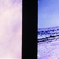
 Probable Choroidal Infarcts After Buckle
-
 Slide 9-23
Feb 26 2019 by Lancaster Course in Ophthalmology
Choroidal infarct (Elschnig spot). There is a localized area of loss of retinal pigment epithelium and outer ischemic retinal atrophy with loss of the photoreceptor cell and outer plexiform layers, and partial loss of the inner nuclear layer without any reparative changes.
Condition/keywords: choroidal infarction, Elschnig's spots
-
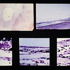
 Slide 9-64
Feb 26 2019 by Lancaster Course in Ophthalmology
Peripheral punched-out lesion from choroidal infarction. The lesion is surrounded by hypertrophic RPE which gives the lesion a dark halo. There is loss of the choriocapillaris, RPE, and outer retinal layers with no reparative proliferation.
Condition/keywords: choroidal infarction, retinal pigment epithelium (RPE) hypertrophy
-
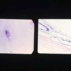
 Slide 9-65
Feb 26 2019 by Lancaster Course in Ophthalmology
Elschnig spot. Localized choroidal infarction with loss of choriocapillaris, RPE, and outer layers of the retina. The thinned inner nuclear layer of the retina rests against Bruch's membrane. At the anterior margin (lower left view) there is an abrupt transition (arrow) between the normal area (left) where the choriocapillaris and RPE are intact and the area of post-ischemic atrophy of the structures (right). A similar but reversed configuration is observed at the posterior margin (lower right view).
Condition/keywords: Bruch's membrane, choroidal infarction, Elschnig's spots
-
 Penetrating Globe Injury
Oct 2 2013 by Jerald A. Bovino, MD
There was a metallic foreign body that caused a rutpured glove. An adjacent vitreous hemorrhage with posterior scleral rupture and adjacent choroidal infarction is present.
Condition/keywords: penetration, ruptured globe, vitreous hemorrhage
 Loading…