Search results (5 results)
-
---thumb.jpg/image-square;max$300,300.ImageHandler) Central Retinal Vascular Obstruction
Central Retinal Vascular Obstruction
Oct 9 2013 by Maurice F. Rabb
Thirty five year old black male with low-grade uveitis. An unusual cause of central retinal vascular obstruction.
Condition/keywords: central retinal vascular obstruction
-
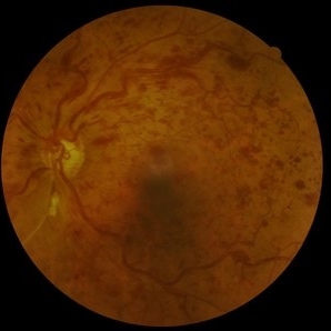 Central retinal vein obstruction (CRVO)
May 29 2022 by Eduardo Javier Pinuer Alvarado
Fundus photograph of a 63 year-old woman with central retinal vein obstruction (CRVO). The patient didn´t notice the decrease of VA, we found this in a routine exam for diabetic patients.
Photographer: Eduardo Pinuer A, Universidad Austral de Chile.
Imaging device: CR-2 AF Digital Non-Mydriatic Retinal Camera, Canon.
Condition/keywords: central retinal vascular obstruction, diabetes, fundus photograph
-
 The Great Vascular Traffic Jam: Combined Retinal Vein and Artery Occlusion
Oct 29 2025 by SHRADDHA RAJ SHRIVASTAVA
57 year old female, recently diagnosed with accelerated hypertension, developed Right eye Combined Retinal Vein and Artery Occlusion. Posterior pole image showed severe disc edema with peri-papillary hemorrhages. There is significant retinal whitening suggestive of edema leading to the classic cherry red spot at the macula. We can also see segmented flow of blood in retinal arterioles, which is the characteristic cattle-trucking seen in central retinal artery occlusion (CRAO). Widefield image revealed multiple intra-retinal blot hemorrhages in all quadrants with tortuous dilated vessels suggestive of central retinal vein occlusion (CRVO).
Photographer: Dr. Shraddha Raj Shrivastava
Imaging device: Nidek Mirante SLO/OCT (Confocal scanning/Spectral domain OCT)
Condition/keywords: central retinal artery occlusion (CRAO), central retinal vascular obstruction, central retinal vein occlusion (CRVO), CRVO with macular edema
-
 The Great Vascular Traffic Jam: Combined Retinal Vein and Artery Occlusion
Oct 29 2025 by SHRADDHA RAJ SHRIVASTAVA
57 year old female, recently diagnosed with accelerated hypertension, developed Right eye Combined Retinal Vein and Artery Occlusion. Posterior pole image showed severe disc edema with peri-papillary haemorrhages. There is significant retinal whitening suggestive of edema leading to the classic cherry red spot at the macula. We can also see segmented flow of blood in retinal arterioles, which is the characteristic cattle-trucking seen in central retinal artery occlusion (CRAO). Widefield image revealed multiple intra-retinal blot hemorrhages in all quadrants with tortuous dilated vessels suggestive of central retinal vein occlusion (CRVO).
Photographer: Dr. Shraddha Raj Shrivastava
Imaging device: Nidek Mirante SLO/OCT (Confocal scanning/Spectral domain OCT)
Condition/keywords: central retinal vascular obstruction, central retinal vein occlusion (CRVO), CRVO with macular edema
 Loading…