Search results (93 results)
-
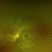
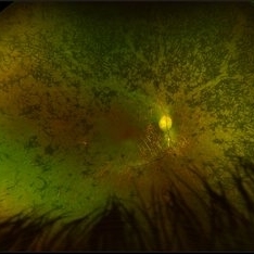
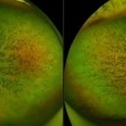 Advanced RP
Nov 5 2024 by rahul saradge
A man, 58, arrived complaining of BOV for both near and distance vision in both eyes, with a 6/9 BCVA in each eye. For a year, the patient had been taking medication for both diabetes and hypertension. In both eyes, the dilated ophthalmoscopic retina revealed waxy disc pallor paired with bony spicules in the mid-periphery. The patient was prescribed spectacles and given counseling regarding the nature of the illness.
Photographer: Lokesh Dukare ,Isha Netralaya Thane
Imaging device: optos
Condition/keywords: bone spicule, optic disc pallor, Optos, Retinitis Pigmentosa
-
---thumb.jpg/image-square;max$300,300.ImageHandler) Bone Spicule
Bone Spicule
Feb 20 2013 by From the Collections of Thomas M. Aaberg, MD and Thomas M. Aaberg Jr., MD
FA and fundus photo of periperal bone spicule
Condition/keywords: bone spicule
-
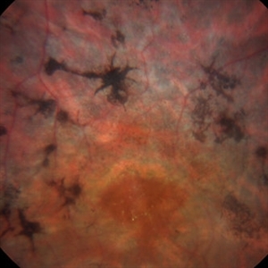
 Bone Spicules
Aug 1 2013 by From the Collections of Thomas M. Aaberg, MD and Thomas M. Aaberg Jr., MD
Bone spicules.
Condition/keywords: bone spicule
-
 Bone Spicules
Mar 1 2014 by Homayoun Tabandeh, MD, FASRS
Bone spicule pigmentary retinopathy in a patient with retinitis pigmentosa.
Condition/keywords: bone spicule, retinitis pigmentosa
-
---thumb.jpg/image-square;max$300,300.ImageHandler) Pattern Dystrophy
Pattern Dystrophy
Aug 12 2013 by From the Collections of Thomas M. Aaberg, MD and Thomas M. Aaberg Jr., MD
Central atrophy with bone spicule.
Condition/keywords: bone spicule, central choroidal atrophy, total, pattern macular dystrophy
-
 Peripheral Bone Spicules
Jul 31 2013 by From the Collections of Thomas M. Aaberg, MD and Thomas M. Aaberg Jr., MD
Peripheral bone spicules.
Condition/keywords: bone spicule, peripheral bone spicules
-
 Peripheral Bone Spicules
Jul 31 2013 by From the Collections of Thomas M. Aaberg, MD and Thomas M. Aaberg Jr., MD
Peripheral bone spicules.
Condition/keywords: bone spicule, peripheral bone spicules
-
 Perivascular Bone Spicule Changes
Mar 1 2021 by Sophia El Hamichi, MD
A 19-year-old female African-American, who is followed for lattice degeneration and bone spicule changes OU. VA 20/20 OU. The bone spicule changes are stable throughout her follow-ups
Condition/keywords: bone spicule, lattice degeneration, Optos, perivascular, white without pressure
-
 Pigmentary Retinal Dystrophy
Mar 29 2019 by Jessica Norkus
Heidelberg Spectralis image of 41-year-old male patient with pigmentary retinal dystrophy. Atypical findings due to unilateral presentation. Patient has been experiencing symptoms for 15 years, notes significant nyctalopia.
Photographer: Jessica Norkus
Imaging device: Heidelberg Spectralis
Condition/keywords: bone spicule, Heidelburg Spectralis, optical coherence tomography (OCT), pigment changes, unilateral blindness
-
 Pigmentary Retinal Dystrophy
Mar 29 2019 by Jessica Norkus
Optos ultra wide field image of 41-year-old male patient with pigmentary retinal dystrophy. Atypical findings due to unilateral presentation. Patient has been experiencing symptoms for 15 years, notes significant nyctalopia.
Photographer: Jessica Norkus
Imaging device: Optos Ultra Wide Field Camera
Condition/keywords: abnormal fundus, bone spicule, color fundus photograph, color photo, fundus autofluorescence (FAF), fundus photograph, Optos, peripheral bone spicules, pigment changes, ultra-wide field imaging, unilateral blindness
-
 Pigmentary Retinal Dystrophy
Mar 29 2019 by Jessica Norkus
Optos ultra wide field image of 41-year-old male patient with pigmentary retinal dystrophy. Atypical findings due to unilateral presentation. Patient has been experiencing symptoms for 15 years, notes significant nyctalopia.
Photographer: Jessica Norkus
Imaging device: Optos Ultra Wide Field Camera
Condition/keywords: abnormal fundus, bone spicule, color fundus photograph, color photo, fundus photograph, Optos, peripheral bone spicules, pigment changes, ultra-wide field imaging, unilateral blindness
-
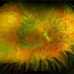 Retinitis Pigmentosa
May 26 2017 by Olivia Rainey
Ultra-wide-field pseudocolor image of the right eye of an 39-year-old female with Retinitis Pigmentosa. She had slightly atypical appearance due to asymmetry: sectoral atrophy in left eye, compared to 360 degree bone spicule formation in right eye. Ddx: Pigmentary degeneration vs infection vs X-linked RP carrier due to asymmetry. Recommended genetic testing through My Retina Tracker, as well as visual field and ERG testing. Patient's vision was sc20/100 PH 20/70 in the right eye and sc20/80 PH 20/40 in the left.
Photographer: Olivia Rainey
Imaging device: Optos California
Condition/keywords: bone spicule, fundus photograph, Optos, peripheral bone spicules, pseudocolor, retinitis pigmentosa, ultra-wide field imaging
-
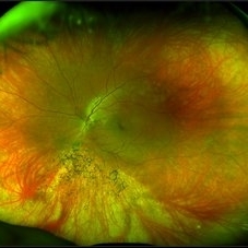 Retinitis Pigmentosa
May 26 2017 by Olivia Rainey
Ultra-wide-field pseudocolor image of the left eye of an 39-year-old female with Retinitis Pigmentosa. She had slightly atypical appearance due to asymmetry: sectoral atrophy in left eye, compared to 360 degree bone spicule formation in right eye. Ddx: Pigmentary degeneration vs infection vs X-linked RP carrier due to asymmetry. Recommended genetic testing through My Retina Tracker, as well as visual field and ERG testing. Patient's vision was sc20/100 PH 20/70 in the right eye and sc20/80 PH 20/40 in the left eye.
Photographer: Olivia Rainey
Imaging device: Optos California
Condition/keywords: bone spicule, fundus photograph, left eye, Optos, peripheral bone spicules, pseudocolor, retinitis pigmentosa, ultra-wide field imaging
-
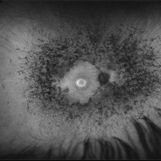 Retinitis Pigmentosa
May 26 2017 by Olivia Rainey
Ultra-wide-field pseudocolor image of the left eye of an 39-year-old female with Retinitis Pigmentosa. She had slightly atypical appearance due to asymmetry: sectoral atrophy in left eye, compared to 360 degree bone spicule formation in right eye. Ddx: Pigmentary degeneration vs infection vs X-linked RP carrier due to asymmetry. Recommended genetic testing through My Retina Tracker, as well as visual field and ERG testing. Patient's vision was sc20/100 PH 20/70 in the right eye and sc20/80 PH 20/40 in the left eye.
Photographer: Olivia Rainey
Imaging device: Optos California
Condition/keywords: autofluorescence imaging, bone spicule, hyperautofluorescent ring, hypoautofluorescence, Optos, peripheral bone spicules, retinitis pigmentosa, ultra-wide field imaging
-
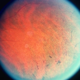
---thumb.jpg/image-square;max$300,300.ImageHandler) Retinitis Pigmentosa
Retinitis Pigmentosa
Feb 20 2013 by From the Collections of Thomas M. Aaberg, MD and Thomas M. Aaberg Jr., MD
peripheral/midperipheral bone spicules
Condition/keywords: bone spicule, retinitis pigmentosa
-
---thumb.jpg/image-square;max$300,300.ImageHandler) Retinitis Pigmentosa
Retinitis Pigmentosa
Feb 20 2013 by From the Collections of Thomas M. Aaberg, MD and Thomas M. Aaberg Jr., MD
peripheral bone spicules
Condition/keywords: bone spicule, retinitis pigmentosa
-
---thumb.jpg/image-square;max$300,300.ImageHandler) Retinitis Pigmentosa
Retinitis Pigmentosa
Feb 20 2013 by From the Collections of Thomas M. Aaberg, MD and Thomas M. Aaberg Jr., MD
bone spicules
Condition/keywords: bone spicule, retinitis pigmentosa
-
---thumb.jpg/image-square;max$300,300.ImageHandler) Retinitis Pigmentosa
Retinitis Pigmentosa
Feb 20 2013 by From the Collections of Thomas M. Aaberg, MD and Thomas M. Aaberg Jr., MD
retinitis pigmentosa
Condition/keywords: bone spicule, retinitis pigmentosa
-
---thumb.jpg/image-square;max$300,300.ImageHandler) Retinitis Pigmentosa
Retinitis Pigmentosa
Feb 20 2013 by From the Collections of Thomas M. Aaberg, MD and Thomas M. Aaberg Jr., MD
peripheral bone spicules
Condition/keywords: bone spicule, retinitis pigmentosa
-
---thumb.jpg/image-square;max$300,300.ImageHandler) retinitis pigmentosa
retinitis pigmentosa
Feb 20 2013 by From the Collections of Thomas M. Aaberg, MD and Thomas M. Aaberg Jr., MD
peripheral bone spicules
Condition/keywords: bone spicule, retinitis pigmentosa
-
---thumb.jpg/image-square;max$300,300.ImageHandler) Retinitis Pigmentosa
Retinitis Pigmentosa
Feb 20 2013 by From the Collections of Thomas M. Aaberg, MD and Thomas M. Aaberg Jr., MD
peripheral bone spicules
Condition/keywords: bone spicule, retinitis pigmentosa
-
---thumb.jpg/image-square;max$300,300.ImageHandler) Retinitis Pigmentosa
Retinitis Pigmentosa
Feb 20 2013 by From the Collections of Thomas M. Aaberg, MD and Thomas M. Aaberg Jr., MD
360 degree bone spicules
Condition/keywords: bone spicule, retinitis pigmentosa
-
---thumb.jpg/image-square;max$300,300.ImageHandler) Retinitis Pigmentosa
Retinitis Pigmentosa
Feb 20 2013 by From the Collections of Thomas M. Aaberg, MD and Thomas M. Aaberg Jr., MD
peripheral bone spicules
Condition/keywords: bone spicule, retinitis pigmentosa
 Loading…