Search results (24 results)
-
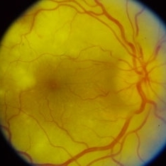
 Hypertensive Retinopathy
Sep 12 2023 by Ben Serar
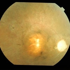
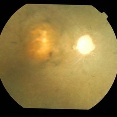
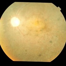
Fundus photograph of LE showing Disc edema with optic disc pallor, hard exudates with dot-blot haemorrhages at the macula ,along with arteriolar attenuation, in a case of Hypertensive retinopathy.
Condition/keywords: arteriolar attenuation, disc edema, Hard exudates, hypertensive retinopathy
-
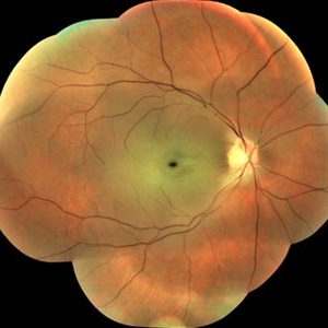
 Hypertensive Retinopathy
Sep 12 2023 by Ben Serar
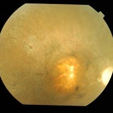
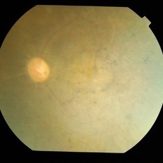
Fundus photograph of RE showing Disc pallor and arteriolar attenuation, with dot-blot and flame-shaped haemorrhages at the posterior pole in a case of Hypertensive Retinopathy.
Condition/keywords: arteriolar attenuation, hypertensive retinopathy, pale disc
-
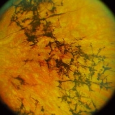 Retinitis Pigmentosa (RP)
Sep 12 2023 by Ben Serar
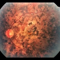
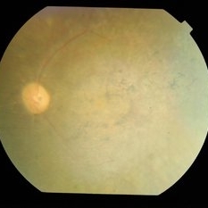
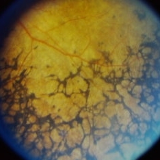
Fundus photograph showing bony spicules and arteriolar attenuation in a case of Retinitis Pigmentosa.
Condition/keywords: arteriolar attenuation, Bony spicules, Retinitis Pigmentosa (RP)
-
 Central Retinal Artery Occlusion
Mar 2 2021 by Renata Garcia Franco, Md
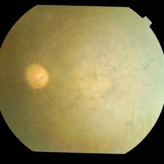
Retinal edema, cherry spot, retinal arteriolar attenuation and segmentation of blood in retinal arterioles.
Photographer: Guillermina Hernandez
Imaging device: Zeiss
Condition/keywords: central artery
-
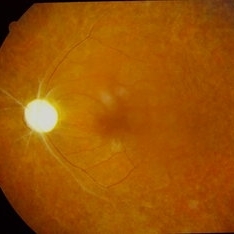
 Central Retinal Artery Occlusion(CRAO) with cilioretinal artery sparing
Sep 12 2023 by Ben Serar
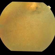
Fundus photograph of RE showing retinal edema and opacification, with normal perfusion at the macula, with arteriolar attenuation and disc edema ,in a case of Central Retinal Artery Occlusion(CRAO) with cilioretinal artery sparing.
Condition/keywords: central retinal artery occlusion (CRAO), cilioretinal artery sparing, disc edema
-
---thumb.jpg/image-square;max$300,300.ImageHandler) late-phase FA showing arteriolar attenuation and late staining of choroidal lesions
late-phase FA showing arteriolar attenuation and late staining of choroidal lesions
Feb 14 2013 by From the Collections of Thomas M. Aaberg, MD and Thomas M. Aaberg Jr., MD
late-phase FA showing arteriolar attenuation and late staining of choroidal lesions
Condition/keywords: multifocal choroiditis, posterior segment inflammation, white dot syndrome
-
 Optic atrophy
Sep 14 2023 by Ben Serar
Fundus photograph of LE showing Pale disc in a case of optic atrophy, with arteriolar attenuation with vascular sheathing and granular fundus.
Condition/keywords: optic atrophy
-
 Proliferative Sickle Cell Retinopathy, Color OD
May 23 2018 by Hosam Attia, MD
45-year-old African American, male with sickle cell anemia (SC disease) with arteriolar attenuation, mild venous tortuosity, Sunburst (S) and large, partially auto-infarcted sea fan with fresh heme, OD.
Imaging device: Optos California Ultra-Wide Field Fundus Camera
Condition/keywords: neovascularization elsewhere (NVE), proliferative retinopathy, sea fan, sickle cell, sickle cell retinopathy
-
 Proliferative Sickle Cell Retinopathy, Color OD
May 23 2018 by Hosam Attia, MD
45-year-old African American, male with sickle cell anemia (SC disease) with arteriolar attenuation, mild venous tortuosity, Sunburst (S) and large, partially auto-infarcted Seafan with fresh heme, OD.
Imaging device: Optos California Ultra-Wide Field Fundus Camera
Condition/keywords: neovascularization elsewhere (NVE), proliferative retinopathy, sea fan, sickle cell, sickle cell retinopathy
-
 Proliferative Sickle Cell Retinopathy, Color OS
May 23 2018 by Hosam Attia, MD
45-year-old African American, male with sickle cell anemia (SC disease ) with arteriolar attenuation, mild venous tortuosity, peripheral arterio-venous anastomoses (shown better on red free), multiple small NVEs/ early sea fans (one w/ early auto-infarction) and sunburst (S) - (Not showing very well in photos) OS.
Imaging device: Optos California Ultra-Wide Field Fundus Camera
Condition/keywords: neovascularization elsewhere (NVE), proliferative retinopathy, sea fan, sickle cell, sickle cell retinopathy
-
 Proliferative Sickle Cell Retinopathy, Red Free OD
May 23 2018 by Hosam Attia, MD
Red free fundus photo of a 45-year-old African American, male with sickle cell anemia (SC Disease ) with arteriolar attenuation, mild venous tortuosity, Sunburst (S) and large, partially auto-infarcted sea fan, OD.
Imaging device: Optos California Ultra-Wide Field Fundus Camera
Condition/keywords: neovascularization elsewhere (NVE), proliferative retinopathy, sea fan, sickle cell, sickle cell retinopathy
-
 Proliferative Sickle Cell Retinopathy, Red Free OS
May 23 2018 by Hosam Attia, MD
Red free fundus photograph of a 45-year-old African American, male with sickle cell anemia (SC disease) with arteriolar attenuation, mild venous tortuosity, peripheral arterio-venous anastomoses (Inferotemporally), multiple small NVEs/ early sea fans OS.
Photographer: Aaron Appiah, M.D.
Imaging device: Optos California Ultra-Wide Field Fundus Camera
Condition/keywords: neovascularization elsewhere (NVE), proliferative retinopathy, sea fan, sickle cell, sickle cell retinopathy
-
 Retinitis Pigmentosa
Apr 6 2022 by Marianna Kavalaraki, MD, Msc
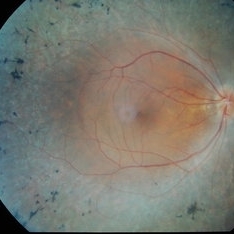
Fundus photography of a 21-year-old man with retinitis pigmentosa. Fundus findings include retinal pigmentary changes in the form of widespread pigment clumpings predominantly in the mid-peripheral fundus, arteriolar attenuation, RPE and retinal atrophy in the posterior pole.
Photographer: Marianna Kavalaraki, General Hospital of Nikaia Piraeus, Department of Ophthalmology
Imaging device: Canon CF-60DSi Digital Fundus Camera
Condition/keywords: retinitis pigmentosa
-
 Retinitis Pigmentosa
Sep 22 2014 by Mallika Goyal, MD
Right fundus of a 32-year-old lady with bilateral retinitis pigmentosa. She has progressive visual complaints starting at age 5, and is the offspring of a consanguineous marriage. Marked disc pallor, retinal arteriolar attenuation, pigment disturbance and macular degeneration are classic features.
Photographer: Mallika Goyal, MD, Apollo Health City, Jubilee Hills, Hyderabad-500033
Condition/keywords: retinitis pigmentosa
-
 Retinitis Pigmentosa
Sep 22 2014 by Mallika Goyal, MD
Inferior retina of a 32-year-old lady with bilateral retinitis pigmentosa. She has progressive visual complaints starting at age 5, and is the offspring of a consanguineous marriage. Marked disc pallor, retinal arteriolar attenuation, pigment disturbance and macular degeneration are classic features.
Photographer: Mallika Goyal, MD, Apollo Health City, Jubilee Hills, Hyderabad-500033
Condition/keywords: retinitis pigmentosa
-
 Retinitis Pigmentosa
Sep 22 2014 by Mallika Goyal, MD
Right fundus of a 32-year-old lady with bilateral retinitis pigmentosa. She has progressive visual complaints starting at age 5, and is the offspring of a consanguineous marriage. Marked disc pallor, retinal arteriolar attenuation, pigment disturbance and macular degeneration are classic features.
Photographer: Mallika Goyal, MD, Apollo Health City, Jubilee Hills, Hyderabad-500033
Condition/keywords: retinitis pigmentosa
-
 Retinitis Pigmentosa
Sep 22 2014 by Mallika Goyal, MD
Left fundus of a 32-year-old lady with bilateral retinitis pigmentosa. She has progressive visual complaints starting at age 5, and is the offspring of a consanguineous marriage. Marked disc pallor, retinal arteriolar attenuation, pigment disturbance and macular degeneration are classic features.
Photographer: Mallika Goyal, MD, Apollo Health City, Jubilee Hills, Hyderabad-500033
Condition/keywords: retinitis pigmentosa
-
 Retinitis Pigmentosa (RP)
Sep 21 2023 by Ben Serar
Fundus photograph showing bony spicules and arteriolar attenuation in a case of Retinitis Pigmentosa.
Condition/keywords: Retinitis Pigmentosa (RP)
-
 Retinitis Pigmentosa (RP)
Sep 14 2023 by Ben Serar
Fundus photograph of the RE showing bony spicules in the mid-periphery, with waxy disc pallor and arteriolar attenuation in a case of retinitis pigmentosa.
Condition/keywords: Retinitis Pigmentosa (RP)
 Loading…