Search results (19 results)
-
 Acute Retinal Detachment
Oct 12 2012 by Jeffrey G. Gross, MD, FASRS
Acute RD with operculated retinal hole.
Condition/keywords: acute retinal detachment, operculated retinal hole
-
 Acute Retinal Detachment
Oct 12 2012 by Jeffrey G. Gross, MD, FASRS
Acute RD with posterior retinal tear, with hemorrhage.
Condition/keywords: acute retinal detachment, posterior retinal tear
-
 Acute Retinal Detachment
Nov 9 2012 by Norman Byer
This 54-year-old man was referred because of sudden symptoms in his opposite eye in which he had suffered an acute retinal detachment secondary to a horseshoe tear around lattice degeneration. During the examination, the fellow eye shown here was also found to have this large horseshoe tear about 1 o’clock hour (4 disc diameters) in size. A tear occurred around a lattice lesion which is present on the flap but is out of focus. This tear had been asymptomatic even though it was caused by a posterior vitreous detachment and illustrates that even very large tears may produce no symptoms or mild symptoms that are easily overlooked.
Condition/keywords: lattice degeneration, posterior vitreous detachment
-
 Acute Retinal Detachment with Posterior Tear
Oct 16 2012 by Jeffrey G. Gross, MD, FASRS
Acute RD with posterior tear.
Condition/keywords: acute retinal detachment, posterior tear
-
 Buckle intrusion with Retinal detachment
Feb 8 2018 by Manish Nagpal, MD, FRCS (UK), FASRS
Patient operated on 10 years back for a scleral buckling surgery presented with decreased vision and had a superonasal retinal detachment along with intrusion of the scleral buckle.
Photographer: Mehul Prajapati
Condition/keywords: acute retinal detachment, retinal break, scleral buckle
-
 GRT Detachment of 10+ Clock Hours With Folded Retina
Sep 11 2025 by Luis J Haddock, MD
Fundus photo of giant retinal tear detachment involving 10+ hours of continuous tearing of the retina, visible anterior edge of retina over GRT.
Photographer: Natella Romero, University of Miami, Bascom Palmer Eye Institute
Imaging device: Optos
Condition/keywords: acute retinal detachment, Giant retinal tear
-
 Horseshoe Tear
Jun 24 2015 by Andree Henaine-Berra, MD
Photograph of the right eye of a 58-year-old male patient with a retinal detachment due to a peripheral horseshoe tear, showing the moment when cryotherapy is applied during the scleral bluckling procedure.
Photographer: Jorge Morales, MD. Hospital General "Dr. Manuel Gea Gonzalez". Mexico City.
Condition/keywords: acute retinal detachment, cryotherapy, scleral buckle
-
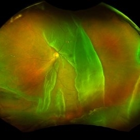
 Macula-Sparing GRT RRD
Jul 6 2017 by Andrew A. Moshfeghi, MD, MBA, FASRS
Wide-field fundus photograph of a 43-year-old myopic man with a history of lattice retinal degeneration status posterior barrier laser performed elsewhere who presented with a giant-retinal tear associated retinal detachment of the right eye.
Photographer: Jay Jiang, University of Southern California Roski Eye Institute
Imaging device: Optos California
Condition/keywords: acute retinal detachment, giant retinal tear, lattice degeneration
-
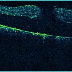
 OCT RRD Macular Off
Jul 9 2014 by Susanna S. Park, MD, PhD
OCT of the macula of a 72-year-old woman with gradual loss of vision from a macular involving inferior rhegmatogenous retinal detachment. Note the corrugated appearance of the retinal elevation.
Photographer: Ellen Redenbo
Condition/keywords: acute retinal detachment, optical coherence tomography (OCT)
-
 PVR Related Recurrent RD
May 19 2014 by John W. Kitchens, MD
Recurrent PVR RD (pre-op photo).
Photographer: Ed SLade
Imaging device: Optos 200Tx
Condition/keywords: acute retinal detachment, proliferative vitreoretinopathy (PVR)
-
 RD Montage
Jul 3 2021 by Somnath Chakraborty, MD
Fundus photo montage of the left eye of a 56-year-old male showing subtotal retinal detachment with macular involvement and a large circumlinear tear extending from 1 o' clock to 3 o' clock hours.
Photographer: Pulak Roy
Condition/keywords: acute retinal detachment, retinal detachment of the macula, retinal tear, retinal tear with detachment
-
 RD With Macular Hole
Nov 27 2020 by Priya Rasipuram Chandrasekaran, MBBS, DO, DNB, FRCS
A 42-year-old female presented with sudden decrease in vision in the right eye and fundus examination showed bullous retinal detachment.
Condition/keywords: acute retinal detachment
-
 Retinal Detachment
Aug 23 2020 by Renata Bertazzi
Fundus photograph of an 76-year-old man with a total retinal detachment.
Photographer: Renata Bertazzi, Instituto Paulista de ensino e Pesquisa em Oftalmologia, São Paulo, SP
Imaging device: DayTona - Optos
Condition/keywords: acute retinal detachment
-
---thumb.jpg/image-square;max$300,300.ImageHandler) Retinal detachment and responsible tear
Retinal detachment and responsible tear
Dec 19 2012 by Eric A. Postel, MD
Stereo color fundus photographs of rhegmatogenous retinal detachment and causative retinal tear
Condition/keywords: acute retinal detachment, bullous retinal detachment, retinal tear
-
 Shafer's Sign
Jan 3 2020 by Manuel Ángel Alcántara Delgado, MD
Slit lamp photograph of a 58-year-old man with rhegmatogenous retinal detachment and tobacco dust presence.
Photographer: Manuel Ángel Alcántara Delgado, CMN SXXI, Mexico City
Condition/keywords: acute retinal detachment, retina surgery, vitrectomy
-
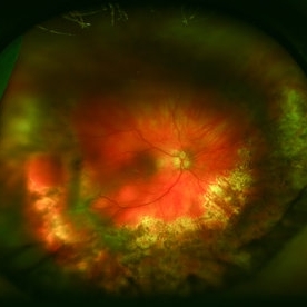
 Total Retinal Detachment
Apr 8 2019 by Gary R. Cook, MD, FACS
63-year-old white female with a total rhegmatogenous retinal detachment OD; macula off; V.A. = 20/200
Imaging device: Topcon VT-50
Condition/keywords: acute retinal detachment
-
 Traumatic Retinal Dialysis-RD
Jan 1 2013 by John T. Thompson, MD
Traumatic retinal dialysis with localized retinal detachment after blunt trauma.
Condition/keywords: acute retinal detachment, retinal dialysis, retinal tear
-
 TRD with Macular Hole
Dec 8 2020 by Priya Rasipuram Chandrasekaran, MBBS, DO, DNB, FRCS
Horizontal 5 line raster scan through the macula shows full thickness macular hole along with separation of neuro sensory retina from the retinal pigment epithelium.
Condition/keywords: acute retinal detachment, traumatic macular hole
-
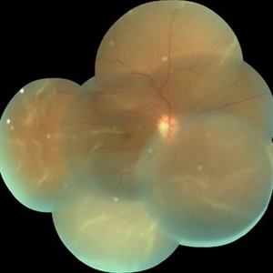
 Ultra-Widefield Image of Retinal Detachment with Giant Retinal Tear-Pre and Post Vitrectomy with Silicone Oil Tamponade
Jan 26 2021 by Kushal S Delhiwala, MBBS, MS, FMRF,FICO, FAICO
Ultra-widefield fundus photograph of a 35-year-old phakic male right eye retinal detachment associated with giant retinal tear from 8 to 1 o' clock hours(left image). Post-op day 1 ultra-widefield image following vitrectomy and silicone oil tamponade (right image) shows reattached retina with fresh laser marks at GRT edge.
Photographer: Kushal Delhiwala, Netralaya superspeciality eye hospital, Ahmedabad, Gujarat,India
Imaging device: Optos Daytona
Condition/keywords: acute retinal detachment, giant retinal tear, pars plana vitrectomy (PPV), perfluorocarbon fluid, perfluorooctane, silicone oil
 Loading…