Search results (18 results)
-
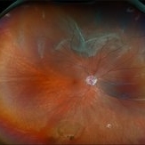
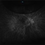
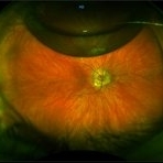
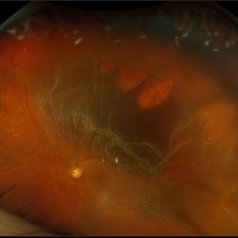
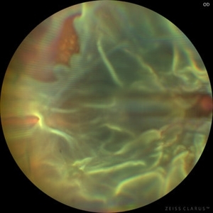 Chronic Open Funnel Retinal Detachment With Horse Shoe Tear
Feb 7 2024 by Harsh Vardhan Singh, MS
67 year old male with history of cataract surgery 1 year presented with old chronic retinal detachment with open funnel configuration with multiple breaks.
Photographer: Harsh Vardhan Singh
Imaging device: Clarus 700
Condition/keywords: chronic retinal detachment, Retinal Detachment, Retinal Detachment with multiple breaks
-
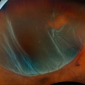
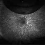
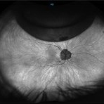
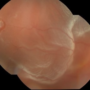 Mac off Retinal Detachment with Horseshoe Tear
Dec 5 2023 by Virginia Gebhart
68 year old male presented with HM vision in OD. Near total detachment with multiple breaks. Scheduled PPV with GFE. Visual prognosis guarded
Photographer: Virginia Gebhart
Imaging device: Topcon
Condition/keywords: Retinal Detachment, retinal detachment of the macula, Retinal Detachment with multiple breaks
-
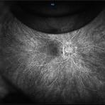
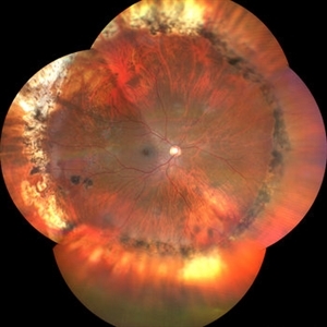
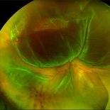 Retinal Detachment with multiple breaks
May 26 2023 by Stephanie M Thurston
Retinal Detachment with multiple breaks; Lattice Degeneration with atrophic holes 54 year old male.
Photographer: Stephanie Thurston
Condition/keywords: Retinal Detachment with multiple breaks
-
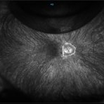
 Retinal Detachment with Multiple Breaks
Aug 12 2025 by Kimberly Wakester
Optomap RGB of a 59-year-old man with a retinal detachment with multiple breaks in the left eye. Surgery was recommended. Patient is to continue follow up care post operatively.
Photographer: Kimberly Wakester, COA, OCT-C, Retina Consultants of Carolina
Imaging device: Optos California
Condition/keywords: lattice degeneration, left eye, Retinal Detachment with Multiple Breaks
-
 Retinal Detachment with Multiple Breaks
Nov 4 2024 by Kimberly Wakester
Ultra-widefield Fundus photograph of an 18-year-old woman with a Retinal detachment with multiple breaks in the right eye. Patient has high Myopia in both eyes. Patient was treated with scleral buckle placement with cryo laser in the right eye and is doing we post operatively.
Photographer: Kimberly Wakester, COA
Imaging device: Optos
-
 Retinal Detachment with Multiple Breaks
Mar 5 2025 by Kimberly Wakester
Optomap RGB image of an 44-year-old man with a retinal detachment with a complex lattice break in the right eye. Surgery was recommended. Patient is to continue follow up care post operatively.
Photographer: Kimberly Wakester, COA
Imaging device: Optos California
Condition/keywords: Retinal Detachment, retinal tear
-
 Retinal Detachment with Multiple Breaks
Feb 3 2025 by Kimberly Wakester
Fundus photograph of a 67-year-old man with a retinal detachment with multiple breaks in the right eye. Patient is doing well s/p PPV and will continued to be observed during PO period.
Photographer: Kimberly Wakester, COA
Imaging device: Optos California
Condition/keywords: horseshoe tear, multiple retinal tears, retinal detachment
-
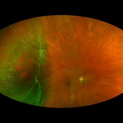 Rhegmatogenous Macula-On Retinal Detachment (Honeycomb)
Aug 6 2024 by Xitlali Caterina
Ultra-wide field fundus photograph of a 72 year old female with a macula-on retinal detachment with multiple breaks affecting her right eye. Patient presented in the office with flashes of light for five consecutive days prior. The patients vision was sc20/30 PHNI. The physician also noted an acute posterior vitreous detachment and lattice degeneration in the affect eye.
Photographer: Xitlali Caterina
Imaging device: Optos California RGB
Condition/keywords: honeycomb, lattice degeneration, Optos, posterior vitreous detachment, Retinal Detachment with Multiple Breaks, rhegmatogenous retinal detachment, ultra-wide field imaging
-
 Gas Bubble
May 21 2021 by Raj K. Maturi, MD
S/P PPV 73-year-old woman with small retinal detachment. Originally presented with decrease vision and residual vitreous floaters. Upon examination small localized retinal detachment with multiple breaks.
Photographer: Charlotte Harris ,Midwest Eye Institute 10300 N Illinois St, Carmel Indiana 46290
Imaging device: Nikon Optos California P200TDx
Condition/keywords: gas bubble
-
 Gas Bubble
May 21 2021 by Raj K. Maturi, MD
S/P PPV 73-year-old woman with small retinal detachment. Originally presented with decrease vision and residual vitreous floaters. Upon examination small localized retinal detachment with multiple breaks.
Photographer: Charlotte Harris ,Midwest Eye Institute 10300 N Illinois St, Carmel Indiana 46290
Imaging device: Nikon Optos California P200TDx
Condition/keywords: gas bubble
-
 Gas Bubble
May 21 2021 by Raj K. Maturi, MD
S/P PPV 73-year-old woman with small retinal detachment. Originally presented with decrease vision and residual vitreous floaters. Upon examination small localized retinal detachment with multiple breaks.
Photographer: Charlotte Harris ,Midwest Eye Institute 10300 N Illinois St, Carmel Indiana 46290
Imaging device: Nikon Optos California P200TDx
-
 Post PPV for retinal detachment
Oct 13 2022 by Vishal Agrawal, MD, FRCS,FACS,FASRS
3 month Post operative picture of right eye .The patient had PPV + gas (C3F8) for total retinal detachment with multiple breaks.
Photographer: Pankaj, Agrawal Hospital,Jaipur
Imaging device: Clarus 700
-
 Repaired Retinal Detachment with Multiple Breaks
Dec 9 2024 by Virginia Gebhart
FAF in 25 year old female of repaired retinal detachment 1.5 year s/p scleral buckle/cryo. Pt had been having symptoms for over a year, inferior demarcation line from retinal fluid that was present. Retina remains flat and attached under buckle. Treated lattice inferiorly, no new holes or tears. VA 20/20
Photographer: Virginia Gebhart, Retina Consultants of Carolina
Imaging device: Optos California
Condition/keywords: autofluorescence imaging, cryotherapy, demarcation line, lattice degeneration, scleral buckle
-
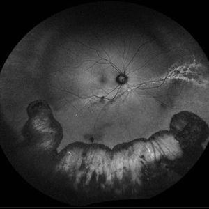
 Retinal Detachment with Multiple OCT Overlays
Jan 7 2025 by Drew Mitchell
Optos 360* Color photo montage with multiple Zeiss Cirrus OCT scan overlays. Retinal Detachment with multiple breaks and a Epiretinal Membrane.
Photographer: Drew Mitchel, OCT-C
Imaging device: Optos California
Condition/keywords: ERM, macular pucker, montage, Optos, OPTOS CALIFORNIA, RD, Retinal Detachment
-
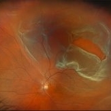
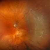
 Rhegmatogenous Macula Off Retinal Detachment with Multiple Breaks
May 29 2024 by Alexis Singstock
Ultra widefield fundus photograph of a 66 year old male with rhegmatogenous macula off retinal detachment with multiple breaks. Patient presented emergently for a curtain/veil in inferonasal visual field. Patient reports the curtain/veil in left eye started about 1 week prior, yet denied seeing flashes and floaters. Patient's vision was hand motion. Dr. Edward Korot examined the patient and scheduled him for a scleral buckle along with pars plana vitrectomy surgery.
Photographer: Alexis Singstock, Retina Specialists of Michigan
Imaging device: Optos California
Condition/keywords: fundus photography, left eye, macula off retinal detachment, OPTOS CALIFORNIA, pars plana vitrectomy (PPV), rhegmatogenous retinal detachment, scleral buckle, ULTRA WIDE FIELD
 Loading…