Search results (1816 results)
-
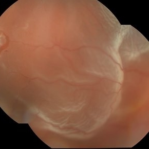
 A Large Break at the Posterior Pole With RD With PVR (S/p Old Blunt Trauma)
Jan 16 2025 by Anand Temkar
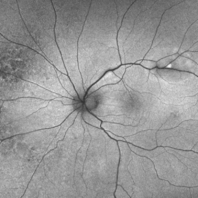
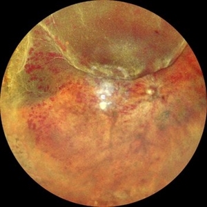
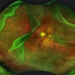
Right eye central fundus color photo of a 10 year old kid who noticed diminution of vision in right eye since a month. We can see the large break at the posterior pole with rolled up margins associated with retinal detachment and PVR changes.
Photographer: Dr.Anand Temkar- Retina Foundation, Ahmedabad
Imaging device: Mirante
Condition/keywords: Posterior pole break, proliferative vitreoretinopathy (PVR), Retinal Detachment
-
 A Large Break at the Posterior Pole With RD With PVR (S/p Old Blunt Trauma)
Jan 16 2025 by Anand Temkar
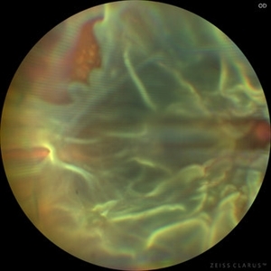
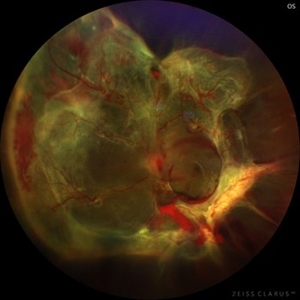
Right eye widefield fundus color photo of a 10 year old kid who noticed diminution of vision in right eye since a month. We can see the large break at the posterior pole with rolled up margins associated with retinal detachment and PVR changes.
Photographer: Dr.Anand Temkar- Retina Foundation, Ahmedabad
Imaging device: Mirante
Condition/keywords: posterior pole break, proliferative vitreoretinopathy (PVR), Retinal Detachment
-
 Blister Retinal Detachment Superotemporal with a Flap Tear
Apr 10 2025 by Daniela Bogenschutz
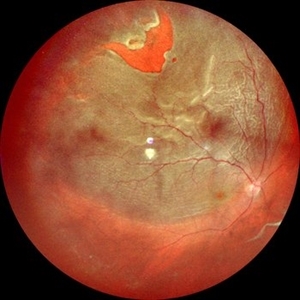
Autofluorescence of a 70-year-old male with a superotemporal retinal detachment prior to having an OCT with unusual findings. Patient states symptoms were "starburst" in his vision in the location of the retinal detachment with the retinal tear. Surgery was scheduled immediately to avoid further progression.
Photographer: Daniela Bogenschutz, OSC; Retina Consultants of the Carolinas, PA
Condition/keywords: Retinal Detachment, retinal detachment with single break
-
 Choroidal Melanoma with Exudative Detachment
Apr 7 2025 by Virginia Gebhart
Autofluorescence image of 36 year old female showing demarcation line of fluid/detachment from new choroidal melanoma. Pt will be scheduled for brachytherapy pending CT scan results.
Photographer: Virginia Gebhart, Retina Consultants of Carolina
Imaging device: Optos California
Condition/keywords: Autoflourescence, autofluorescence imaging, choroidal melanoma, melanoma, retinal detachment
-
 Choroidal Melanoma with Exudative Retinal Detachment
Mar 2 2023 by Aditya S Kelkar, MS, FRCS, FASRS,FRCOphth
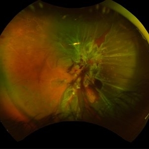
Color fundus photograph of the left eye of a 45 year old male showing choroidal melanoma with exudative retinal detachment.
Photographer: Dr. Pranali Surawase, National Institute of Ophthalmology, Pune, India.
Imaging device: Zeiss Clarus 500
Condition/keywords: choroidal mass, exudative retinal detachment, Retinal detachment
-
 Chronic Open Funnel Retinal Detachment With Horse Shoe Tear
Feb 7 2024 by Harsh Vardhan Singh, MS
67 year old male with history of cataract surgery 1 year presented with old chronic retinal detachment with open funnel configuration with multiple breaks.
Photographer: Harsh Vardhan Singh
Imaging device: Clarus 700
Condition/keywords: chronic retinal detachment, Retinal Detachment, Retinal Detachment with multiple breaks
-
 Chronic RD with Retinal Dialysis
Jul 23 2025 by Virginia Gebhart
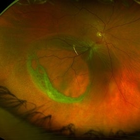
64 year old female with chronic retinal detachment from head trauma 41 years ago. Peripheral scarring from 6:00 to 11:00 with area of subretinal fluid inferotemporally, well demarcated with subretinal bands. Retinal dialysis inferotemporal from 7:00 to 9:00. No surgical repair needed or recommended at this time.
Photographer: Virginia Gebhart, Retina Consultants of Carolina
Imaging device: Optos California
Condition/keywords: chronic retinal detachment, demarcation, RD, Retinal Detachment, retinal dialysis, subretinal bands
-
 Combined Traction and Rhegmatogenous Retinal Detachment From Proliferative Diabetic Retinopathy
Mar 27 2025 by Nikhil K Bommakanti, MD
A middle-aged patient presented with a combined traction and rhegmatogenous retinal detachment.
Condition/keywords: Active PDR Tractional retinal Detachment, PDR, Retinal Detachment, rrd, TRD
-
 Complex Retinal Detachment with PVR and Starfold
Jun 6 2025 by Jenn Geelan
57 year old male with a Complex Retinoschisis related retinal detachment with PVR and a Posterior Star Fold
Photographer: Jenn Geelan, Retina-Vitreous Surgeons of CNY
Imaging device: Optos California
Condition/keywords: proliferative vitreoretinopathy (PVR), rare, Retinal Detachment, retinoschisis, Starfolds, subretinal fluid
-
 CRVO with RD
Apr 4 2025 by Tejaswita Verma
Fundus photo of a 58 year-old hypertensive male who presented with RE 6/60 vision with CRVO, rhegmatogenous RD.H/O DOV in RE since 3 days, H/O receiving anti VEGF X 2 injections 2 months ago
Photographer: DR. TEJASWITA VERMA
Imaging device: MIRANTE
Condition/keywords: central retinal vein occlusion (CRVO), Retinal Detachment
-
 Diabetic traction retinal detachment
Jan 9 2023 by JORGE SOBERANES
Proliferative diabetic retinopathy with extensive traction retinal detachment in a patient with type 1 diabetes mellitus.
Photographer: Dr. Jorge I. Soberanes, Asociación para Evitar la Ceguera en México.
Imaging device: Zeiss Clarus 700
Condition/keywords: Retinal Detachment, tractional retinal detachment
-
 Dislocation of the Crystalline Lens with a Retinal Detachment
Apr 21 2025 by Hrishikesh Naik, MS
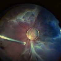
An intraoperative screen grab shows a dislocation of the crystalline lens along with an associated rhegmatogenous retinal detachment in a case of Marfan’s syndrome. The case was managed by a combined PPV-SB procedure. A vitrectomy cutter is seen at the left.
Photographer: Hrishikesh Naik
Condition/keywords: intraoperative, lens dislocation, Marfan's syndrome, Retinal Detachment, vitrectomy
-
 Giant Retinal Tear
Feb 20 2024 by Soobien Lee
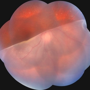
Optos color fundus photograph of a 40-year-old caucasian male who is a UFC fighter with a total retinal detachment in his right eye secondary to a giant retinal tear from 10 o'clock to 2 o'clock.
Photographer: Trinity Wolf, Elman Retina Group
Imaging device: Optos Ultra-Widefield Imaging
Condition/keywords: giant retinal tear, optos, Retinal Detachment, Retinal tear with detachment, trauma
-
 Giant Retinal Tear
Jul 15 2024 by Arthi Mohankumar , MS,MRCS ED, FICO,FAICO
Fundus montage of a 15 year old boy with Marfans syndrome who presented with defective vision in the right eye.
Photographer: Arthi Mohankumar
Condition/keywords: giant retinal tear, Retinal detachment
-
 Giant Retinal Tear
Oct 13 2022 by Vishal Agrawal, MD, FRCS,FACS,FASRS
65 year old female presented with sudden vision loss. On examination superior giant tear with retinal detachment was noted in the right eye.
Photographer: Mahipal,Agrawal Hospital
Imaging device: Clarus 700
Condition/keywords: giant retinal tear, Retinal Detachment
-
 Giant Retinal Tear with Bare Choroid
Dec 13 2024 by Thirumalesh Mochi Basavaraj, MD
Intra-operative view of a Pediatric Giant Retinal Tear with a view of the Bare Choroid Superiorly.
Photographer: Thirumalesh Mochi Basavaraj
Imaging device: Leica Proveo 8
Condition/keywords: GIANT RETINAL TEAR, Retinal Detachment
-
 Giant Retinal Tear with Multiple Retinal Breaks
Apr 21 2025 by Hrishikesh Naik, MS
A 28 year old high myope with retinal detachment associated with a supero-temporal giant retinal tear in addition to multiple peripheral horseshoe tears and an additional supero-nasal retinal tear.
Photographer: Hrishikesh Naik
Imaging device: Optos Daytona
Condition/keywords: giant retinal tear, High Myopia, horseshoe tear, retinal break, retinal detachment
-
 Hemorrhagic Choroidals
Jan 22 2025 by Danish Shabbir, Ophthalmic Technologist
78 year old female complains of suddenly vision decrease 2 days ago.
Photographer: Danish Shabbir,Retina-EyeCare Centre
Imaging device: Optos California
Condition/keywords: choroidal detachment, Retinal Detachment, retinal detachment with choroidal
-
 Horse Shoe Tear With Retinal Detachment
Apr 28 2025 by rohan jain
56 year-old male with idiopathic HST and RRD
Photographer: Dr. ROHAN JAIN
Condition/keywords: horseshoe tear, Retinal Detachment, rrd
-
 Inadvert Globe Perfuration After Peribulbar Block
Mar 13 2025 by Bruno B Ribeiro
Fundus photograph of a 74-year-old woman who underwent pars plana vitrectomy OS due to rhegmatogenous retinal detachment. A horseshoe retinal tear can be seen at 5h. Intraoperative evaluation revealed a chorioretinal scar with the shape of the needle track at the same location. Despite rare, globe perfuration after peri or retrobulbar block may happen, even by the most experienced anesthesiologist.
Photographer: Bruno Barbosa Ribeiro, Angelina Meireles
Imaging device: Optos California
Condition/keywords: retinal detachment
-
 Intra-operative still of PFCL bubble in RD Surgery
Apr 15 2023 by Veer Singh, MS, FVRS, FMRF, FICO (Retina)
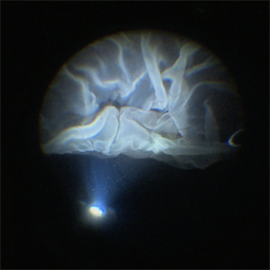
Intra-operative still of PFCL bubble in RD Surgery. PFCL being used to flatten the detached retina.
Photographer: Dr. Veer Singh
Condition/keywords: intraoperative, pars plana vitrectomy (PPV), PFCL, Retinal Detachment
-
 Intraoperative View of a Giant Retinal Tear
Dec 13 2024 by Thirumalesh Mochi Basavaraj, MD
Intraoperative view of 12 year old child with Giant retinal tear with Retinal detachment.
Photographer: Thirumalesh Mochi Basavaraj
Imaging device: Lumera Proveo 8
Condition/keywords: GIANT RETINAL TEAR, PVR, Retinal Detachment
-
 Long-standing RD with PVR
Jan 28 2022 by Gayathri Mohan
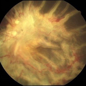
Color fundus photograph showing a long standing RD with PVR with fixed retinal folds.
Photographer: Dr Gayathri Mohan
Imaging device: Canon
Condition/keywords: PVR, Retina Folds, Retinal Detachment
-
 Mac off Retinal Detachment with Horseshoe Tear
Dec 5 2023 by Virginia Gebhart
68 year old male presented with HM vision in OD. Near total detachment with multiple breaks. Scheduled PPV with GFE. Visual prognosis guarded
Photographer: Virginia Gebhart
Imaging device: Topcon
Condition/keywords: Retinal Detachment, retinal detachment of the macula, Retinal Detachment with multiple breaks
-
 Mac-on Retinal Detachment (Barely!)
Feb 6 2025 by Virginia Gebhart
FAF of 46 year old male with a mac-on retinal detachment from 1:00 to 6:00 with a single break at 3:00. Pt scheduled for emergent PPV/Laser/GFE
Photographer: Virginia Gebhart, Retina Consultants of Carolina
Imaging device: Optos California
Condition/keywords: autofluorescence imaging, retinal detachment
 Loading…