Search results (15 results)
-
 RAM
RAM
Jun 25 2024 by Tejaswita Verma
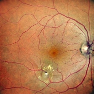
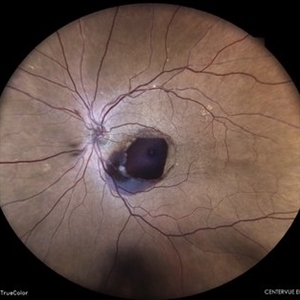
Right eye Fundus photo of a 73 year old female with 6/ 9 vision having retinal artery macroaneurysm.
Photographer: DR. TEJASWITA VERMA
Imaging device: MIRANTE
Condition/keywords: RETINAL ARTERY MACROANEURYSM
-
 Retinal Artery Macroaneurysm
Jul 13 2024 by Tejaswita Verma
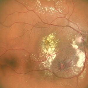
A 53 year old female presented with blurred vision in RE since a month ,with borderline DM and HTN not on medications .H/o highest BP recording was 160/90 mm Hg.Vision 6/60 .FFA revealed leakages. She was advised RE focal laser with intravitreal anti-VEGF injections
Photographer: DR. TEJASWITA VERMA
Imaging device: MIRANTE
Condition/keywords: RETINAL ARTERY MACROANEURYSM
-
 Retinal Artery Macroaneurysm
Sep 29 2019 by Haider Ali
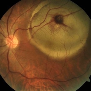
65-year-old male with progressively decreasing vision in his left eye for last three months.
Photographer: Dr Haider Ali Chaudhry, Madinah Teaching Hospital
Imaging device: Retinal Artery Macroaneurysm
Condition/keywords: arteriolar macroaneurysm, hypertensive retinopathy, retinal arterial macroaneurysm
-
 Retinal Artery Macroaneurysm
May 2 2014 by Neha Goel, MS DNB FRCS (Glasg)
Fundus photograph of a 60-year-old hypertensive male who presented with sudden diminution of vision in his left eye since 10 days.
Photographer: Neha Goel
Imaging device: Zeiss visucam
Condition/keywords: retinal arterial macroaneurysm, subretinal hemorrhage
-
 Retinal Artery Macroaneurysm FFA
May 2 2014 by Neha Goel, MS DNB FRCS (Glasg)
Fluorescein angiogram of the patient showing a retinal artery macroaneurysm and blocked fluorescence from the subretinal hemorrhage.
Photographer: Neha Goel
Imaging device: Zeiss Visucam
Condition/keywords: retinal arterial macroaneurysm, subretinal hemorrhage
-
 Early Phase Angiogram Showing Vitreous, Preretinal and Subretinal Hemorrhage
Aug 10 2014 by Thomas A. Ciulla, MD, MBA, FASRS
A 90-year-old woman presented with sudden loss of central vision OS of 1 week. Her VA was finger counting OS, with mild-moderate vitreous hemorrhage and large preretinal and subretinal hemorrhage, felt to be due to AMD or retinal artery macroaneurysm.
Condition/keywords: submacular hemorrhage, tissue plasminogen activator (tPA), vitrectomy, wet age-related macular degeneration (wet AMD)
-
 Laser Induced BRAO in IRVAN Syndrome
May 3 2019 by Deependra Vikram Singh, MD FASRS
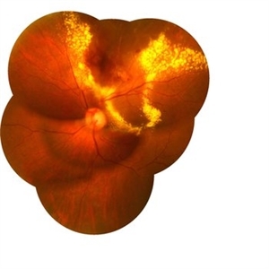
Fundus photograph of a 26-year-old man with IRVAN syndrome referred for vitreous surgery in OS for secondary rhegmatogenous retinal detachment. OD has received laser photocoagulation for capillary nonperfusion areas and retinal artery macroaneurysm associated with retinal vasculitis. Fundus photograph of OD shows laser induced nasal BRAO. Case re-emphasizes why laser for macroaneurysm should be avoided in cases with IRVAN.
Photographer: Deependra V Singh, Eye-Q Superspecialty Eye Hospitals. Gurugram, India
Imaging device: Zeiss Visucam 500
Condition/keywords: arteriolar macroaneurysm, branch retinal artery occlusion (BRAO), laser photocoagulation
-
 Late Phase Angiogram Showing Vitreous, Preretinal and Subretinal Hemorrhage
Aug 10 2014 by Thomas A. Ciulla, MD, MBA, FASRS
A 90-year-old woman presented with sudden loss of central vision OS of 1 week. Her VA was finger counting OS, with mild-moderate vitreous hemorrhage and large preretinal and subretinal hemorrhage, felt to be due to AMD or retinal artery macroaneurysm.
Condition/keywords: submacular hemorrhage, tissue plasminogen activator (tPA), vitrectomy, wet age-related macular degeneration (wet AMD)
-
 Post Vitrectomy Early Phase Angiogram
Aug 10 2014 by Thomas A. Ciulla, MD, MBA, FASRS
She underwent pars plana vitrectomy, submacular tissue plasminogen activator injection, gas injection, face down positioning. Follow up 3 months later showed VA: 20/80 OS. Angiography shows resolution of submacular hemorrhage, no active CNVM, and no retinal artery macroaneurysm.
Condition/keywords: post-vitrectomy, submacular hemorrhage, tissue plasminogen activator (tPA), wet age-related macular degeneration (wet AMD)
-
 Post Vitrectomy Late Phase Angiogram
Aug 10 2014 by Thomas A. Ciulla, MD, MBA, FASRS
She underwent pars plana vitrectomy, submacular tissue plasminogen activator injection, gas injection, face down positioning. Follow up 3 months later showed VA: 20/80 OS. Angiography shows resolution of submacular hemorrhage, no active CNVM, and no retinal artery macroaneurysm.
Condition/keywords: post-vitrectomy, submacular hemorrhage, tissue plasminogen activator (tPA), wet age-related macular degeneration (wet AMD)
-
 Pre-Op Photos Showing Vitreous, Preretinal and Subretinal Hemorrhage
Aug 10 2014 by Thomas A. Ciulla, MD, MBA, FASRS
A 90-year-old woman presented with sudden loss of central vision OS of 1 week. Her VA was finger counting OS, with mild-moderate vitreous hemorrhage and large preretinal and subretinal hemorrhage, felt to be due to AMD or retinal artery macroaneurysm.
Condition/keywords: submacular hemorrhage, tissue plasminogen activator (tPA), vitrectomy, wet age-related macular degeneration (wet AMD)
-
 RAM With Garland of Hard Exudate
Mar 3 2020 by KRISHNENDU NANDI, MS
Fundus Photo of left eye of 75-year-old female with retinal artery macroaneurysm at superior quadrant with garland like hard exudates.
Photographer: KRISHNENDU NANDI
Imaging device: Topcon
Condition/keywords: hard exudates, retinal arterial macroaneurysm
-
 Retinal Macroaneurysm
May 7 2024 by Akansha Sharma
Color fundus photograph of a 74 year old female with retinal artery macroaneurysm.
Photographer: Dr. Akansha Sharma, Bharati Eye Hospital
Condition/keywords: macroaneurysm, RAM
-
 Ruptured retinal artery macroaneurysm
Sep 6 2023 by PRATIK SHENOY, MBBS, DNB, FVRS
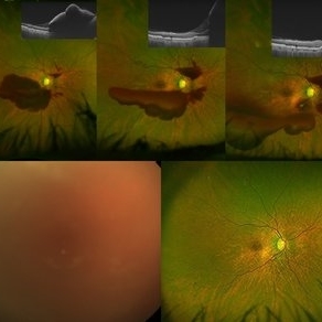
A 66-year-old female presented with a ruptured retinal artery macroaneurysm and a visual acuity of finger counting close to face. The multi layered hemorrhage receded on its own inferiorly with an improvement of visual acuity. However, the patient developed a breakthrough bleed with vitreous hemorrhage three weeks later with a drop in visual acuity to hand movements. She underwent pars plana vitrectomy for the same with an improvement in visual acuity to 6/9.
Photographer: Gaurav Kamble, Isha Netralaya
Imaging device: Optos
Condition/keywords: OCT, Optos, pars plana vitrectomy (PPV), retinal arterial macroaneurysm, vitreous hemorrhage
-
 Ruptured Retinal Artery Macroaneurysm
Jun 18 2024 by KANWALJEET HARJOT MADAN, M.S. (Ophthalmology); FAICO (Vitreous - Retina)
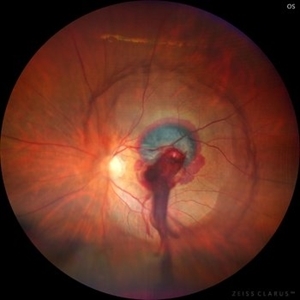
This is a fundus photo depicting ruptured Retinal Artery Macroaneurysm (RAM) in the left eye of a 63 years old female. RAM is an acquired saccular or fusiform dilatation of the retinal arterioles that usually occur within the first three orders of bifurcation. The Superotemporal artery is the most common location. RAM may be asymptomatic or cause a number of complications such as macular edema, serous macular detachment, and hemorrhages.
Photographer: Dr Kanwaljeet Harjot Madan
Condition/keywords: Haemorrhage, macroaneurysm, retinal arteriole
 Loading…