Search results (4 results)
-
 Choroidal Tuberculoma Presenting in the Third Trimester of Pregnancy
May 24 2019 by Unnati Vishwanath Shukla, M. S. ,DNB, FVRS FNERF, MNAMS,PhD Scholar(Retina)
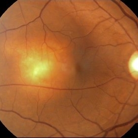
A 29-year-old pregnant female patient (in the third trimester of pregnancy) presented with the complaints of blurring of vision in the right eye and metamorphosia since 1 month. On examination a solitary yellowish elevated subretinal mass of about 2 disc diameters lesion was noted in the right eye temporal to the macula with minimal subretinal fluid around the lesion. Left eye findings were normal. Further investigations revealed normal Chest X- ray and abdominal USG and negative sputum AFB analysis. Thorough uveitic profile was done to rule out other infective and immune causes of choroidal lesion . Positive findings revealed positive mantoux test , positive interferon gamma release essay (Quantiferon Gold). Further aqueous tapping was done which showed presence of acid fast bacilli. Final diagnosis of right eye isolated choroidal tuberculoma was made and appropriate treatment was initiated.
Photographer: Unnati Shukla, C.H. Nagri Eye Hospital, NHL medical college, Ahmedabad,Gujarat,India.
Condition/keywords: choroidal tuberculoma, macula lesion, mantoux test, pregnancy, Quantiferon gold
-
 Central Retinal Vein Occlusion
Jan 21 2022 by Olivia Rainey
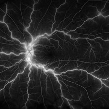
Ultra-widefield fluorescein angiogram of a 23-year-old female with a Central Retinal Vein Occlusion affecting her left eye. The patient presented on 12/22/2021 cc20/40-2 vision in the left eye. The patient reported recent trauma of being hit with a fist on both sides of face followed by vision loss. The patient has history of Hashimoto's thyroid disease. The following labs have been ordered, PT, PTT, CBC, antithrombin III activity, protein C, protein S, Factor V Leiden mutation, Prothrombin (G20210A), lipid panel, HbA1c, quantiferon gold, RPR, and CXR.
Photographer: Olivia Rainey, OCT-C, COA
Imaging device: Optos California
Condition/keywords: central retinal vein occlusion (CRVO), disc leakage, fluorescein angiogram (FA), fluorescein leakage, left eye, non-ischemic central retinal vein occlusion (CRVO), Optos, trauma, ultra-wide field imaging
-
 Serpiginious Chorioretinitis
Sep 6 2023 by PUSHPANJALI BADOLE
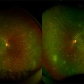
Fundus photograph of an 18 year-old male with bilateral choroiditis. Tuberculosis work-up revealed positive QuantiFERON Gold test and Mantoux test. Patient was started anti-tuberculosis treatment along with oral corticosteroids. Optos fundus photography shows extensive active plus healed lesions with pigmentary change in the midperipheral retina and periphery suggesting varied stage presentation of lesions. There are few hemorrhages in right eye superotemporal retina.
Photographer: Hitesh Rawlani, Isha Netralaya, Kalyan.
Condition/keywords: serpiginous choroiditis
-
 Tuberculoma
Oct 15 2016 by Apoorva Guruprasad Ayachit, MS
Fundus photograph of the left eye of a 28-year-old male who came with DOV OS since 9 days. Mantoux 20 mm induration and Quantiferon gold positive. Responded to anti tubercular treatment.
Photographer: Apoorva Ayachit
Condition/keywords: choroidal tuberculoma
 Loading…