Search results (3 results)
-
 Ehlers-Danlos Syndrome
Apr 22 2021 by Harita Shah
Fundus photograph of a 37-year-old male, known case of Ehlers-Danlos Syndrome, having right eye CNVM scar with angioid streaks.
Photographer: Harita Shah, Banker's Retina Clinic & Laser Centre
Imaging device: Topcon TRC 50DX
Condition/keywords: Ehlers-Danlos syndrome
-
 Ehlers-Danlos Syndrome
Apr 22 2021 by Harita Shah
Fundus photograph of a 37-year-old male, known case of Ehlers-Danlos Syndrome, having left eye CNVM scar with angioid streaks.
Photographer: Harita Shah, Banker's Retina Clinic & Laser Centre
Imaging device: Topcon TRC 50DX
Condition/keywords: Ehlers-Danlos syndrome
-
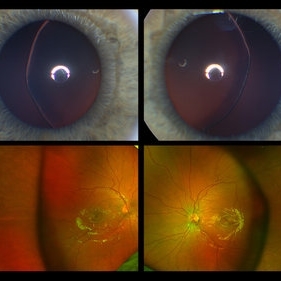 Ectopia Lentis
Jan 21 2021 by Jamin S. Brown, MD
This image serial demonstrates a patient with simple ectopia lentis. Anterior segment photographs in the upper panel demonstrate nasally subluxated crystalline lenses. Widefield fundus photography shows a "pseudo-buckle" which is the result of an optical effect due to the lens subluxation (artifactual image enlargement). Also note the juvenile macular reflex in this young patient. Ectopia lentis can present isolated ("simple") or in combination with various systemic defects (Marfan's syndrome, Weil-Marchesani syndrome or Ehlers-Danlos syndrome to name a few). Isolated ectopia lentis can be hereditary and causative genes have been identified as ADAMTSL4 located on chromosome 4 and FBN1 gene located on chromosome 15. Defects in the genes cause weakness in the zonular fibers which can lead to lens dislocation. Lastly, various ocular disorders such as Aniridia, Axenfeld-Rieger, Pseudoexfoliation or Trauma may also result in lens dislocation or subluxation.
Photographer: Stefanie Palmer CRA, Retina Vitreous Surgeons of CNY
Condition/keywords: dislocated lens, ectopia lentis
 Loading…