Search results (182 results)
-
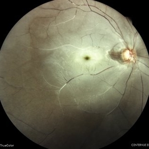
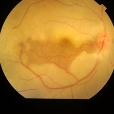 Central Artery Occlusion
Aug 6 2023 by Anjana Mirajkar, MS Ophthalmology
Color photo of a 42 year old male in a case of central artery occlusion with cilio retinal artery sparing
Photographer: Dr. Anjana Mirajkar -Retina Foundation, Ahmedabad
Condition/keywords: Central Retinal Artery Occlusion, central retinal artery occlusion (CRAO)
-
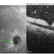 Central Artery Occlusion with Cilio Retinal Artery Sparing
Aug 6 2023 by Anjana Mirajkar, MS Ophthalmology
OCT image (horizontal scan ) of RE of a 42 year old male in a case of central artery occlusion with cilio retinal artery sparing loss of differentiation of retinal layers in nasal half.
Photographer: Dr. Anjana Mirajkar -Retina Foundation, Ahmedabad
Condition/keywords: Central Retinal Artery Occlusion
-
 Central Retinal Artery Occlusion
Nov 15 2022 by T. P . VIGNESH, MBBS,MS
SD-OCT of a 30 year old man with sudden loss of vision in left eye, revealing hyperreflective inner retinal layers .
Photographer: Priyanka
Imaging device: Heidelberg Spectralis
Condition/keywords: Central Retinal Artery Occlusion
-
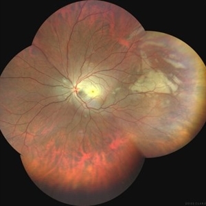 Central Retinal Artery Occlusion
Mar 21 2025 by T. P . VIGNESH, MBBS,MS
Fundus photo montage of a 29 year old man with Central retinal artery occlusion.
Photographer: Bharathi
Imaging device: Zeiss Clarus
Condition/keywords: central retinal artery occlusion (CRAO)
-
 Central Retinal Artery Occlusion
Mar 11 2024 by Dr.Pavithra Subramanian
A 51 year old male with defective vision in right eye for past 4days.On examination RE RAPD present and Fundus examination found to be Right eye Central retinal artery occlusion with Grade 4 Hypertensive Retinopathy.
Photographer: Dr Pavithra Subramanian
Condition/keywords: central retinal artery occlusion (CRAO), malignant hypertension
-
 Central Retinal Artery Occlusion
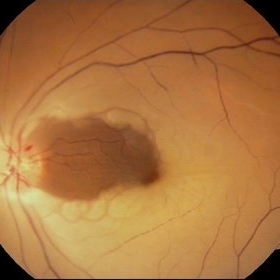
Apr 10 2024 by Tejaswita Verma
Left eye fundus photo of a 75 year old male with pale edematous retina with cherry red spot in a case of central retinal artery occlusion.
Photographer: DR. TEJASWITA VERMA
Imaging device: MIRANTE
Condition/keywords: central retinal artery occlusion (CRAO), cherry red spot
-
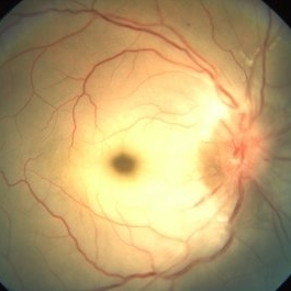
 Central Retinal Artery Occlusion
May 26 2025 by yao zhang
Fundus photograph of an 84-year-old man with CRAO
Photographer: Yao Zhang,TongUniversity, Shanghai East Hospital, Department of ophthalmology
Condition/keywords: Ciliary artery sparing central retinal artery occlusion (CRAO)
-
 Central Retinal Artery Occlusion
Sep 25 2024 by Gustavo Uriel Fonseca Aguirre
43 year-old female with a history of rheumatoid arthritis, a cherry-red spot was observed in the fundus due to central retinal artery occlusion of 8 hours' duration.
Photographer: Gustavo U. Fonseca Aguirre, Fundación Hospital Nuestra Señora de la Luz, Ciudad de México
Condition/keywords: central retinal artery occlusion
-
 Central Retinal Artery Occlusion
Apr 20 2018 by Kim Barrett
64-year-old female woke with no vision in her right eye. This image was taken at 6:11 minutes and the vessels have not filled. Patient has been treated with PRP laser and anti-VEGF therapy. Current vision is CF @ 2 ft.
Photographer: Kim Barrett C.O.A.
Imaging device: Heidelberg
Condition/keywords: central retinal artery occlusion (CRAO), diabetes, hypertension, smoker, uncontrolled
-
 Central Retinal Artery Occlusion
Oct 25 2017 by satar Baghrizabehi
Fundus photograph of an 67-year-old man with uncontrolled arterial hypertension.
Photographer: Satar Baghrizabehi MD Educ. Hospital Rakican
Condition/keywords: central retinal artery occlusion (CRAO)
-
 Central Retinal Artery Occlusion
Feb 20 2013 by From the Collections of Thomas M. Aaberg, MD and Thomas M. Aaberg Jr., MD
No history; documented over time.
Condition/keywords: central retinal artery occlusion (CRAO)
-
 Central Retinal Artery Occlusion
Jun 4 2019 by Unnati Vishwanath Shukla, M. S. ,DNB, FVRS FNERF, MNAMS,PhD Scholar(Retina)
A young female patient of Indian origin on Oral Contraceptive medication presenting with Central Retinal Artery Occlusion with Cilioretinal artery Sparing.
Photographer: Unnati Shukla, C.H. Nagri Eye Hospital, NHL medical college, Ahmedabad,Gujarat,India.
Condition/keywords: central retinal artery occlusion (CRAO), cherry red spot, cilioretinal sparing, pale retina
-
 Central Retinal Artery Occlusion
Jan 13 2020 by Prithvi Chandrakanth
37-years-old male with complaints of sudden diminution of vision in the left eye for the past three days. Fundus examination revealed pale retina in the left eye with cherry red spot and normal fundus picture in right eye.
Photographer: DR.PRITHVI CHANDRAKANTH, ARAVIND EYE HOSPITAL, UDUMALPET
Imaging device: TRASH TO TREASURE RETCAM
Condition/keywords: central retinal artery occlusion (CRAO), cherry red spot, retcam, smartphone fundus photography
-
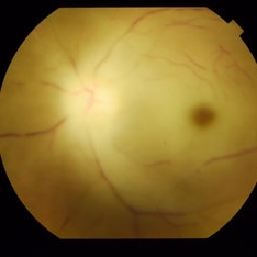
 Central Retinal Artery Occlusion
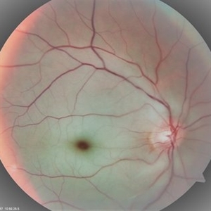
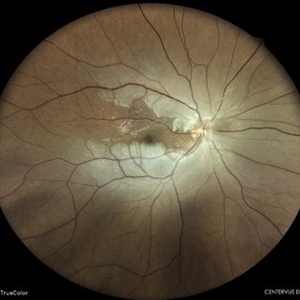
May 7 2024 by Akansha Sharma
Color fundus photograph of a 40 year old female with central retinal artery occlusion.
Photographer: Dr. Akansha Sharma, Bharati Eye Hospital
Condition/keywords: central retinal artery occlusion (CRAO), CRAO
-
 Central Retinal Artery Occlusion
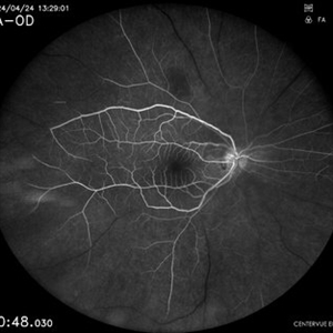
May 7 2024 by Akansha Sharma
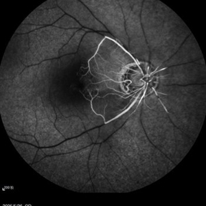
Early phase fluorescein angiography of a 40 year old female with central retinal artery occlusion.
Photographer: Dr. Akansha Sharma, Bharati Eye Hospital
Condition/keywords: central retinal artery occlusion (CRAO), CRAO
-
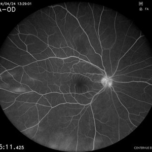 Central Retinal Artery Occlusion
May 7 2024 by Akansha Sharma
Late phase fluorescein angiography of a 40 year old female with central retinal artery occlusion.
Photographer: Dr. Akansha Sharma, Bharati Eye Hospital
Condition/keywords: central retinal artery occlusion (CRAO), CRAO
-
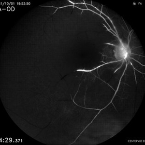 Central Retinal Artery Occlusion
Apr 17 2024 by Akansha Sharma
Fluorescein angiography of a 48 year old male with central retinal artery occlusion.
Photographer: Dr. Akansha Sharma, Bharati Eye Hospital
Condition/keywords: central retinal artery occlusion (CRAO), CRAO
-
 Central Retinal Artery Occlusion
Apr 17 2024 by Akansha Sharma
Color fundus photograph of a 48 year old male with central retinal artery occlusion.
Photographer: Dr. Akansha Sharma, Bharati Eye Hospital
Condition/keywords: central retinal artery occlusion (CRAO), CRAO
-
 Central retinal artery occlusion
Nov 30 2022 by Ethan K Sobol, MD
A central retinal artery occlusion with cilioretinal artery sparing, imaged using a Volk Panretinal 2.2 and an iPhone camera in the emergency department.
Photographer: Jared Raabe, MD, Emory University Hospital
Imaging device: IPhone 13 Pro
Condition/keywords: central retinal artery occlusion (CRAO)
-
---thumb.JPG/image-square;max$300,300.ImageHandler) Central retinal artery occlusion
Central retinal artery occlusion
Oct 26 2012 by Mallika Goyal, MD
Fundus photograph of a 55-year-old gentleman one day after sudden vision loss. Shows retinal infarct with "cherry red" spot at macular centre.
Condition/keywords: central retinal artery occlusion (CRAO), cherry red spot
-
 Central Retinal Artery Occlusion
Aug 28 2018 by Gabriela Lopezcarasa Hernandez, MD
CRAO related to systemic LUE.
Photographer: MARCO ANTONIO SAUZA CASTILLEJOS M.D., MEXICO.
Condition/keywords: central retinal artery occlusion (CRAO), lupus
-
 Central Retinal Artery Occlusion
Aug 28 2018 by Gabriela Lopezcarasa Hernandez, MD
CRAO related to systemic LUES.
Photographer: MARCO ANTONIO SAUZA CASTILLEJOS M.D., MEXICO.
Condition/keywords: central retinal artery occlusion (CRAO)
-
 Central Retinal Artery Occlusion
Aug 28 2018 by Gabriela Lopezcarasa Hernandez, MD
35-year-old women with CRAO and vasculitis due to systemic lupus.
Photographer: MARCO ANTONIO SAUZA CASTILLEJOS M.D., MEXICO.
Condition/keywords: central retinal artery occlusion (CRAO), vasculitis
-
 Central Retinal Artery Occlusion
Aug 28 2018 by Gabriela Lopezcarasa Hernandez, MD
35-year-old women with CRAO and vasculitis due to systemic lupus.
Photographer: MARCO ANTONIO SAUZA CASTILLEJOS M.D., MEXICO.
Condition/keywords: central retinal artery occlusion (CRAO), vasculitis
-
 Central Retinal Artery Occlusion
Apr 22 2021 by Alay S. Banker, MD
Fundus Photograph of a 45-year-old male patient with central retinal artery occlusion in left eye since 14/04/2021. H/O COVID-19 positive RT PCR on 22/03/2021 and COVID -19 negative on 08/04/2021. H/O Post COVID viral pneumonitis and patient was on oxygen 5 Lit/Day. K/C/O DM Type-II
Photographer: Alay S Banker, Banker's Retina Clinic & Laser Centre
Imaging device: Topcon TRC 50DX
Condition/keywords: central retinal artery occlusion (CRAO)
 Loading…