Search results (11 results)
-
 Vitreous Cyst
Jul 13 2013 by Jason S. Calhoun
Patient with a large floater in the left eye. Fundus shows larger vitreous cyst floating in the vitreous. Patient deferred surgery.
Photographer: Jason S. Calhoun, Department of Ophthalmology, Mayo Clinic Jacksonville, Florida
Imaging device: TOPCON TRC 50-EX
Condition/keywords: vitreous, vitreous cyst
-
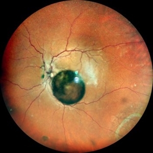
 Congenital Vitreous Cyst
May 1 2018 by G. Robert Hampton, MD
Image of a 42-year-old man with asymptomatic congenital vitreous cyst. This was found on routine exam, he had no idea it was present. Vision 20/20
Photographer: Stefanie Palmer
Imaging device: TRC-50EX
Condition/keywords: vitreous cavity
-
 Vitreous Cyst
Apr 29 2021 by William G. Campbell, MD
Left wide-field fundus photograph of a 50-year-old male with normal visual acuity, but who has always been aware of a clear round circle in his vision.
Photographer: Marina Pascoe, Melbourne Retina Associates, Victoria, Australia
Condition/keywords: congenital anomaly
-
 Pigmented Vitreous Cyst
May 25 2021 by Anmol Naik
A 56-year-old Indian male, known case of panuveitis, presented with complaint of floater in his left eye. On examination, a pigmented floating vitreous cyst was seen in the anterior vitreous. Vitreous cyst, an extremely rare entity, can be congenital or acquired. Detailed examination is necessary to rule out malignancy and parasitic infections.
Photographer: Dr. Anmol Naik, MS, Nakshatra Superspeciality Eye Hospital, Pune, India
Imaging device: Appasamy Associates Slit Lamp AIA-11 3S L model, Chennai, India
Condition/keywords: vitreous cyst
-
 Vitreous Cyst
Oct 16 2022 by Pramod Kumar Suman, MBBS, MD
Fundus photograph of an 63-year-old male with a floating vitreous cyst.
Photographer: Pramod Kumar Suman, Retina Foundation, Ahmedabad
Imaging device: Mirante
Condition/keywords: vitreous cyst
-
 Eye Blossoms
Jul 22 2021 by Vishal Gupta, MBBS, MS
Fundus image of a floating encapsulated vitreous cyst in a 42-year-old diabetic woman resembled a Flower Bud and was doubted to be a hydatid cyst, but was finally confirmed to be an encapsulate vitreous hemorrhage.
Photographer: Dr Vishal Gupta, INHS Asvini, Mumbai, INDIA
Imaging device: Zeiss
Condition/keywords: hemorrhage, vitreous
-
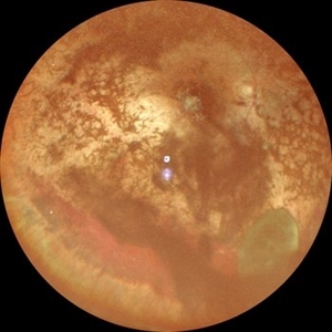
 Universe within the eye
Jul 25 2023 by Pravin Jain, Mbbs, Dnb, FVRS
Fundus photograph of an 25 year old male with a floating vitreous cyst.
Photographer: Dr Sunil Khandelwal
Condition/keywords: vitreous cyst
-
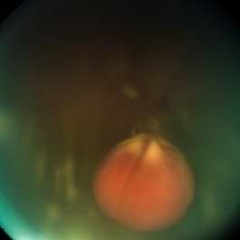
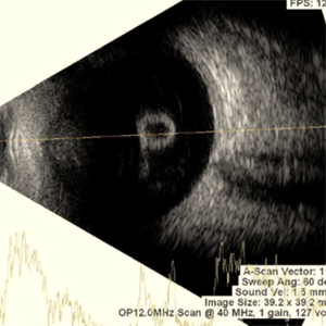 Vitreous Cyst With Vitreous Hemorrhage in a Case of Retinitis Pigmentosa
Oct 5 2024 by Anand Temkar
This picture shows RE USG of a 30 year old female with vitreous cyst with spontaneous vitreous hemorrhage in a case of retinitis pigmentosa.
Photographer: Dr.Anand Temkar- Retina Foundation, Ahmedabad
Condition/keywords: retinitis pigmentosa, vitreous cyst, Vitreous hemorrhage
-
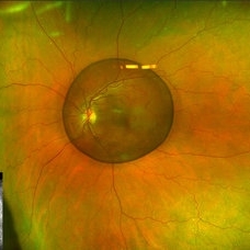
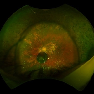 Vitreous Cysts and Asteroid Hyalosis
May 22 2024 by José Laércio Araújo Filho
Fundus Photograph of a elderly patient with retinitis pigmentosa, asteroid hyalosis and a unilateral vitreous cyst.
Photographer: José Laércio de Araújo Filho, Universidade de São Paulo, Brazil
Imaging device: Optos Daytona P200T / A10600
Condition/keywords: asteroid hyalosis, vitreous cyst
-
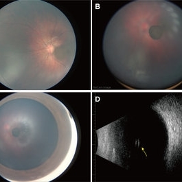 Oval Pigmented Vitreous Cyst
Nov 27 2024 by Xinyu Zhao
An 8-month-old infant was found to have a brown object in the left vitreous during a fundus screening. A wide-field digital retinal camera (RetCam) revealed a pigmented, non-transparent, freely floating, oval cystic lesion in the vitreous, measuring 2 disc diameters (Figures A-D). The cyst appeared cloudy when focused on the retina (Figure A) but was clearly defined in the vitreous (Figure B). Ultrasound showed a well-defined hyperreflective structure with a hyporeflective lumen (Figure D, indicated by the yellow arrow). A diagnosis of a vitreous pigment cyst, rare in infants, was made. Long-term follow-up is necessary to monitor changes affecting the infant’s vision.
Photographer: Xinyu Zhao, Shenzhen Eye Hospital, Shenzhen, China
Imaging device: RetCam
Condition/keywords: infant, vitreous cyst
-
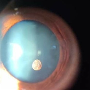
 Vitreous Cyst With Vitreous Hemorrhage in a Case of Retinitis Pigmentosa
Oct 5 2024 by Anand Temkar
This picture shows RE color photo of a 30 year old female with vitreous cyst with spontaneous vitreous hemorrhage in a case of Retinitis Pigmentosa.
Photographer: Dr.Anand Temkar- Retina Foundation, Ahmedabad
Condition/keywords: vitreous cyst, vitreous hemorrhage
 Loading…