Search results (43 results)
-
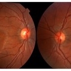
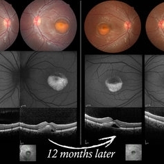 Vitelliform Macular Dystrophy or Best Disease
Dec 16 2016 by Young Hee Yoon, MD, PhD
Bilateral fundus photographs and autofluorescence images of 15-year-old girl who was diagnosed as vitelliform macular dystrophy or Best disease. Vitelliform macular lesion showed morphologic change during one year.
Photographer: Hyejin Jo, Sunghyun Kim, Heoni Hong, Minjung Chae, Mihwa Shin, Asan medical center, Seoul
Imaging device: Topcon TRC-500X fundus camera, Heidelberg HRA 2 autofluorescence, Heldelberg Spectralis OCT
Condition/keywords: Best disease, pseudohypopyon, scrambled-egg, vitelliform macular dystrophy
-
---thumb.jpg/image-square;max$300,300.ImageHandler) Adult Vitelliform Dystrophy
Adult Vitelliform Dystrophy
Feb 13 2013 by From the Collections of Thomas M. Aaberg, MD and Thomas M. Aaberg Jr., MD
Right eye.
Condition/keywords: vitelliform macular dystrophy
-
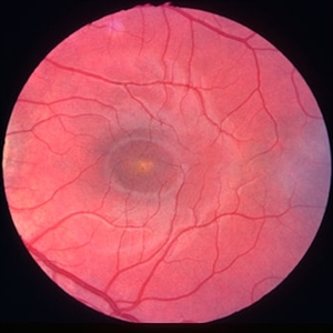
 Vitelliform Macular Dystrophy
Sep 2 2012 by Hyung-Woo Kwak, MD
The typical appearance is of bilateral, round or oval, yellow, symmetrical, subretinal lesions, typically one-third to one-half optic disc diameter in size.
Imaging device: Zeiss F450 plus
Condition/keywords: Best disease
-
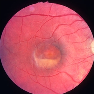
 Best Vitelliform Macular Dystrophy
Jan 14 2018 by Koushik Tripathy, MBBS, MD
Typical subretinal egg yolk appearance of Best disease. Arden ratio in electrooculogram was reduced in either eye.
Condition/keywords: Best disease, vitelliform macular dystrophy
-
---thumb.jpg/image-square;max$300,300.ImageHandler) Adult Vitelliform Dystrophy
Adult Vitelliform Dystrophy
Apr 1 2013 by Henry J. Kaplan, MD
Fundus photograph of a middle aged patient with mild decreased vision and bilateral macular vitelliform lesion #1.
Condition/keywords: adult vitelliform dystrophy, vitelliform lesion, vitelliform macular dystrophy
-
 Best Disease
Oct 12 2012 by Gregg T. Kokame, MD, MMM, FASRS
Best disease
Photographer: Jaclyn Pisano, Retina Consultants of Hawaii
Imaging device: Zeiss FF-450 plus
Condition/keywords: Best disease, vitelliform macular dystrophy
-
---thumb.jpg/image-square;max$300,300.ImageHandler) Adult Vitelliform Dystrophy
Adult Vitelliform Dystrophy
Feb 13 2013 by From the Collections of Thomas M. Aaberg, MD and Thomas M. Aaberg Jr., MD
Left eye
Condition/keywords: left eye, vitelliform macular dystrophy
-
---thumb.jpg/image-square;max$300,300.ImageHandler) Adult Vitelliform Dystrophy
Adult Vitelliform Dystrophy
Mar 29 2013 by Henry J. Kaplan, MD
Fundus photograph of the same patient, left eye #2 notice the multiple vitelliform lesions.
Condition/keywords: vitelliform macular dystrophy
-
 Best disease left eye
Jan 11 2013 by Alex P. Hunyor, MD
Best's vitelliform macular dystrophy, left eye - early vitelliform stage.
Condition/keywords: Best disease
-
---thumb.jpg/image-square;max$300,300.ImageHandler) Adult Vitelliform Dystrophy
Adult Vitelliform Dystrophy
Feb 13 2013 by From the Collections of Thomas M. Aaberg, MD and Thomas M. Aaberg Jr., MD
Right eye.
Condition/keywords: vitelliform macular dystrophy
-
---thumb.jpg/image-square;max$300,300.ImageHandler) Adult Vitelliform Dystrophy
Adult Vitelliform Dystrophy
Feb 13 2013 by From the Collections of Thomas M. Aaberg, MD and Thomas M. Aaberg Jr., MD
Right eye.
Condition/keywords: vitelliform macular dystrophy
-
 Best disease, right eye
Jan 11 2013 by Alex P. Hunyor, MD
Best's vitelliform macular dystrophy, right eye - vitelliruptive stage.
Condition/keywords: Best disease
-
 Best Disease
Jul 5 2014 by John S. King, MD
Late 30s.
Photographer: URMC
Condition/keywords: Best disease, vitelliform macular dystrophy
-
 Best Disease
Jul 5 2014 by John S. King, MD
20s.
Photographer: URMC
Condition/keywords: Best disease, vitelliform macular dystrophy
-
 Best Disease
Jul 5 2014 by John S. King, MD
30s.
Photographer: URMC
Condition/keywords: Best disease, vitelliform macular dystrophy
-
 Best Disease
Jul 5 2014 by John S. King, MD
Teens.
Photographer: URMC
Condition/keywords: Best disease, vitelliform macular dystrophy
-
 Vitelliform Macular Dystrophy
Sep 2 2012 by Hyung-Woo Kwak, MD
The typical appearance is of bilateral, round or oval, yellow, symmetrical, subretinal lesions, typically one-third to one-half optic disc diameter in size.
Imaging device: Zeiss F450 plus
-
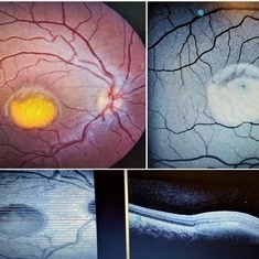
 Vitelliform Macular Dystrophy (VMD) or Best Disease
Apr 22 2018 by Ronald Silva
Fundus photograph and OCT of a 3-year-old boy with Best Disease, and cystoid macular edema (CME) in vitelliform macular dystrophy (VMD) in right eye.
Photographer: Ronald Rocha da Silva, HCOE, Feira de Santana-BA
Imaging device: And OCT
Condition/keywords: Best disease, cystoid macular edema (CME), vitelliform macular dystrophy
-
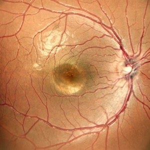
 Best's Vitelliform Macular Dystrophy
Apr 8 2019 by Gary R. Cook, MD, FACS
Vitelliform lesion in the left eye of a 10-year-old female demonstrating a yellow, round, egg yolk-like lesion; V. A. = 20/25
Condition/keywords: Best disease, vitelliform lesion, vitelliform macular dystrophy
-
 Best Vitelliform Macular Dystrophy
Mar 17 2020 by Sophia El Hamichi, MD
Classic "egg yolk" presentation in a 16-year-old female with best disease.
Condition/keywords: autofluorescence imaging, Best disease, optical coherence tomography (OCT), vitelliform macular dystrophy
-
 Best Disease
May 1 2018 by Mitzy E Torres Soriano, MD
Fundus photographs of an 5-year-old boy with best vitelliform macular dystrophy and family history.
Photographer: Luciana García,MD
Condition/keywords: Best disease, vitelliform macular dystrophy
-
 Best Vitelliform Dystrophy with Secondary CNVM
Jan 8 2019 by Rutul R Patel, MD Ophthalmology
10-year-old girl with b/l Best vitelliform dystrophy and left eye secondary CNVM.
Photographer: Dr. Rutul Patel
Imaging device: TOPCON TRC 50DX
Condition/keywords: Best disease, choroidal neovascular membrane (CNVM), vitelliform macular dystrophy
-
 Best Vitelliform Macular Dystrophy
Dec 10 2020 by McGill University Health Centre
Postmortem eyes from 101-year-old female. Past clinical history includes a poor vision for many years due to macular degeneration. The last Visual acuity test recorded 6/15 OD and 6/6 OS. IOP 14 and 18 torr OS. Histopathology: Disclosed and yellow 2x2mm macular lesion. Microscopic examination: elevated placoid macular lesion with overlying pigment granules. Electron microscopy examination: pigment granules with abundant lipofuscin and melanolysosomes, photoreceptor cells markedly attenuated (less degenerated at the periphery) Numerous calcified drusen throughout the retina particularly in the posterior pole. RPE lipofuscin content is elevated in Best’s dystrophy. The extractability of the PRE lipofuscin fluorophores is reduced (it is normal during senescence). The defect in Best’s dystrophy accelerates this age related change in lipofuscin.
Condition/keywords: Best vitelliform macular dystrophy (BVMD), histopathology, pathology
-
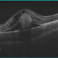 Cystoid Macular Edema (CME) in Vitelliform Macular Dystrophy (VMD)
Apr 22 2018 by Ronald Silva
Macula OCT of a 3-year-old boy with low vision and cystoid macular edema (CME) in vitelliform macular dystrophy (VMD) in right eye.
Photographer: Ronald Rocha da Silva, HCOE, Feira de Santana-BA
Condition/keywords: Best disease, cystoid macular edema (CME), vitelliform macular dystrophy
-
 Best Vitelliform Macular Dystrophy
Jun 26 2022 by Vaidehi Sathaye
8 yr old male child with Best Vitelliform Macular Dystrophy
Photographer: Dr. Vaidehi Sathaye
Imaging device: Mirante
Condition/keywords: Best vitelliform macular dystrophy (BVMD)
 Loading…