Search results (15 results)
-
 Venous Stasis Retinopathy
Feb 20 2015 by H. Michael Lambert, MD
Scattered hemorrhages associated with venous stasis retinopathy in eye with ipsilateral total internal carotid artery occlusion. Vision is 20/40.
Condition/keywords: arterial occlusion, venous stasis retinopathy
-
---thumb.jpg/image-square;max$300,300.ImageHandler) Carotid Cavernous Fistula
Carotid Cavernous Fistula
Jul 29 2013 by Hamid Ahmadieh, MD
Color fundus photograph of the right eye of a 40-year-old man with venous stasis retinopathy secondary to carotid cavernous fistula.
Condition/keywords: carotid-cavernous fistula, venous stasis retinopathy
-
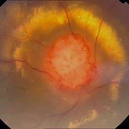 Color Photo of Optic Disc Capillary Hemangioblastoma
Mar 18 2014 by Arwa Azmeh, MD, PhD
Color fundus photograph of an 48-year-old male who complained of decreased visual acuity in his right eye over the last few months. Systemically the patient was healthy. His VA was OD Cf 3m, OS 20/20. Anterior segments were WNL in OU. IOP was WNL in OU. Fundus exam OD revealed unpigmented mass over the optic disc with retinal venous tortuosity at its edges with a ring of thick HYE surrounding it and shallow RD in this area extending to the foveal area. Several few small retinal hemorrhages were seen in the far retinal periphery which were explained to be caused by venous stasis due the optic disc tumor.
Condition/keywords: color photo, optic disc, retinal hemangioblastoma
-
 Optic Nerve Congestion / Venous Stasis
Feb 20 2013 by From the Collections of Thomas M. Aaberg, MD and Thomas M. Aaberg Jr., MD
No history but this set is associated with disc drusen slides.
Condition/keywords: optic nerve drusen, venous stasis
-
 Venous Stasis Retinopathy
Feb 20 2015 by H. Michael Lambert, MD
Scattered hemorrhages associated with venous stasis retinopathy in eye with ipsilateral total internal carotid artery occlusion. Vision is 20/40.
Condition/keywords: arterial occlusion, venous stasis retinopathy
-
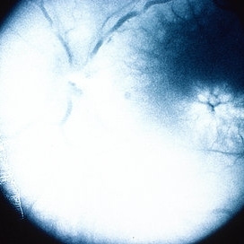
 Venous Stasis Retinopathy
Feb 20 2015 by H. Michael Lambert, MD
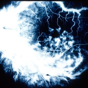
53 second fluorescein angiogram of eye with venous stasis retinopathy in eye with ipsilateral total internal carotid artery occlusion. Fluorescein angiogram shows slow fill. Vision is 20/40.
Condition/keywords: arterial occlusion, venous stasis retinopathy
-
 Venous Stasis Retinopathy
Feb 20 2015 by H. Michael Lambert, MD
Scattered hemorrhages associated with venous stasis retinopathy in eye with ipsilateral total internal carotid artery occlusion. Vision is 20/40.
Condition/keywords: arterial occlusion, venous stasis retinopathy
-
 Venous Stasis Retinopathy
Feb 20 2015 by H. Michael Lambert, MD
45 second fluorescein angiogram of eye with venous stasis retinopathy in eye with ipsilateral total internal carotid artery occlusion. Fluorescein angiogram shows slow fill. Vision is 20/40.
Condition/keywords: venous stasis retinopathy
-
 Venous Stasis Retinopathy
Feb 20 2015 by H. Michael Lambert, MD
68 second fluorescein angiogram of eye with venous stasis retinopathy in eye with ipsilateral total internal carotid artery occlusion. Fluorescein angiogram shows slow fill. Vision is 20/40.
Condition/keywords: arterial occlusion, venous stasis retinopathy
-
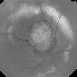 Red Free Photo of Optic Disc Capillary Hemangioblastoma
Mar 18 2014 by Arwa Azmeh, MD, PhD
Red free fundus photograph of an 48-year-old male who complained of decreased visual acuity in his right eye over the last few months. Systemically the patient was healthy. His VA was OD Cf 3m, OS 20/20. Anterior segments were WNL in OU. IOP was WNL in OU. Fundus exam OD revealed unpigmented mass over the optic disc with retinal venous tortuosity at its edges with a ring of thick HYE surrounding it and shallow RD in this area extending to the foveal area. Several few small retinal hemorrhages were seen in the far retinal periphery which were explained to be caused by venous stasis due to the optic disc tumor
Condition/keywords: optic disc, red-free, retinal hemangioblastoma
-
 Optic Nerve Congestion / Venous Stasis
Feb 20 2013 by From the Collections of Thomas M. Aaberg, MD and Thomas M. Aaberg Jr., MD
No history but this set is associated with disc drusen slides; fluorescein angiogram showing leakage.
Condition/keywords: optic nerve drusen, venous stasis
-
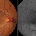 Branch Retinal Artery Occlusion With Central Retinal Vein Occlusion
Jun 19 2019 by Unnati Vishwanath Shukla, M. S. ,DNB, FVRS FNERF, MNAMS,PhD Scholar(Retina)
A 65-year-old male hypertensive patient having resolving nonischemic central retinal vein occlusion presenting with inferotemporal branch retinal artery occlusion.
Photographer: Unnati Shukla
Condition/keywords: branch retinal artery occlusion (BRAO), central retinal vein occlusion (CRVO), cotton wool exudates, venous stasis
-
 Optic Nerve Congestion / Venous Stasis
Feb 20 2013 by From the Collections of Thomas M. Aaberg, MD and Thomas M. Aaberg Jr., MD
No history but this set is associated with disc drusen slides; fluorescein angiogram showing leakage.
Condition/keywords: optic nerve drusen, venous stasis
-
 Venous Stasis Retinopathy
Jun 27 2024 by Akansha Sharma
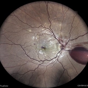
Color fundus photograph of a 31 year old male patient with venous stasis retinopathy.
Photographer: Dr. Akansha Sharma, Bharati Eye Hospital
Condition/keywords: cystoid macular edema (CME), Disc Edema, SHH, Sub hyaloid haemorrhage
-
 Venous Stasis Retinopathy
Aug 6 2024 by Akansha Sharma
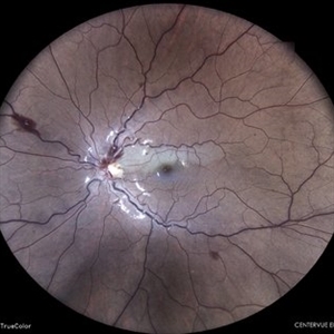
Color fundus photograph of a 18 year old male with venous stasis retinopathy.
Photographer: Dr. Akansha Sharma, Bharati Eye Hospital
Condition/keywords: arterial occlusion, venous stasis retinopathy
 Loading…