Search results (53 results)
-
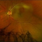
 Preretinal Vascular Loop
Feb 20 2015 by H. Michael Lambert, MD
Vascular loop on the optic nerve. 31-year-old black female, else normal eye exam. Father, brother and daughter also had vascular abnormalities.
Condition/keywords: dominantly inherited, optic nerve, vascular loop
-
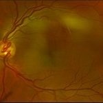
 Congenital prepapillary vascular loop
Jan 11 2013 by Alex P. Hunyor, MD
Congenital prepapillary vascular loop.
Condition/keywords: congenital prepapillary vascular loop
-
 Prepapillary Vascular Loop
Feb 20 2013 by From the Collections of Thomas M. Aaberg, MD and Thomas M. Aaberg Jr., MD
No history or color photo. Flurescein angiogram.
Condition/keywords: prepapillary vascular loop
-
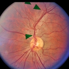
 Congenital Prepapillary Arterial Loop With a Figure-of-Eight Configuration
Mar 27 2019 by Tammy Mclaughlin
Congenital prepapillary arterial loop with a figure-of-eight configuration. OD. There is a twisted anomalous vessel eminating from the optic disc into the vitreous. Likely a congenital anomaly. Does not require treatment and should not be a vision threat.
Photographer: Tammy Mclaughlin, Carolina Retina Center 645 W. Wesmark Blvd. Sumter, Sc 29150
Condition/keywords: congenital anomaly, congenital prepapillary vascular loop
-
 Preretinal Vascular Loop
Feb 20 2015 by H. Michael Lambert, MD
Vascular loop on the optic nerve. 31-year-old black female, else normal eye exam. Father, brother and daughter also had vascular abnormalities.
Condition/keywords: dominantly inherited, optic nerve
-
 Vascular loops in retinopathy of prematurity
Nov 3 2013 by Maria Ana Martinez-Castellanos, MD
Angiography of a baby with ROP treated with intravitreal anti-angiogenic therapy 1 week prior to the time this photo was taken. We can see the active vascular remodeling, vascular loops, the place where the demarcation line was at the time of the diagnosis and the growth of new vessels into the avascular zone, the leakage corresponds to immature vessels not fully covered by mural cells and not due to an inflammatory reaction.
Photographer: Maria A. Martinez-Castellanos. Asociacion para Evitar la Ceguera en Mexico
Imaging device: RetCam II
Condition/keywords: anti-VEGF, retinopathy of prematurity (ROP)
-
 Preretinal Vascular Loop
Feb 20 2015 by H. Michael Lambert, MD
Vascular loop on the optic nerve. 29-year-old black male, else normal eye exam. Father, sister and niece also had vascular abnormalities.
Condition/keywords: dominantly inherited, optic nerve, vascular loop
-
 Autosomal Dominant Preretinal Vascular Loops
Jan 5 2015 by H. Michael Lambert, MD
Color photo showing corkscrew loop on right optic nerve.
Condition/keywords: vascular loop
-
 Vascular Loops
Feb 20 2015 by H. Michael Lambert, MD
Vascular Loop, optic nerve right eye. Adult black male.
Condition/keywords: prepapillary vascular loop
-
 Vascular Loops
Feb 20 2015 by H. Michael Lambert, MD
Inferior artery off the optic nerve loops around the vein.
Condition/keywords: vascular loop
-
 Preretinal Vascular Loop
Feb 20 2015 by H. Michael Lambert, MD
Vascular loop on the optic nerve. 31-year-old black female, else normal eye exam. Father, brother and daughter also had vascular abnormalities.
Condition/keywords: dominantly inherited, optic nerve, vascular loop
-
 Vascular Loop
Jul 11 2013 by Jerald A. Bovino, MD
No history, slide is labeled vascular loop, this is the same slide, or stereo pair to #475.
Condition/keywords: vascular loop
-
 Peri-papillary Vascular Loop
Jun 2 2020 by Dhaivat Shah
Peri-papillary vascular loops (PVL) are rare congenital vascular malformations, which are usually detected as accidental finding during routine fundus examination. They can often be confused with tributary vein occlusion or racemose hemangioma. Although benign and asymptomatic, they can be rarely associated with vitreous hemorrhage and arterial occlusion. We herein present a case of a 60-year-old hypertensive male, who was diagnosed elsewhere to have a tributary vein occlusion and was referred to us. FFA was advised to rule out neovascularization, surrounding capillary non perfusion and mass lesion (hemangioma). On FFA, the arterial loop showed a slightly delayed filling (3-5 seconds) as compared to the other arterial vessels and the original vessel appeared to be a branch arising from central retinal artery. The choroidal filling was delayed in the area supplied by the loop. A cilioretinal artery was also noted. The patient was diagnosed to have a Peri-papillary vascular arterial loop (PVL), likely to be congenital in origin. The patient was reassured and was advised yearly follow up. These loops are usually accidental findings discovered during routine fundus examination. Since these vessels are looped and tortuous, they exhibit a slower and laminar blood flow, which make them more prone for arterial occlusions. The vitreous in this area tends to be adherently attached, so during PVD induction, it is likely to cause a tear and hemorrhage leading to vitreous hemorrhage. Until and unless there is a break, this hemorrhage tends to resolve on its own and does not warrant treatment. If there is an evident break, it can be dealt with laser barrage.
Photographer: Choithram Netralaya
Condition/keywords: congenital prepapillary vascular loop
-
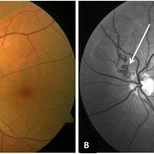
 Prepapillary Vascular Loop
Mar 11 2020 by Asdrubal F Moreno, MD
Fundus color photograph of a 80-year-old woman with a unilateral congenital prepapillary vascular loop and hypertensive retinopathy, focused on the retinal plane for perception.
Photographer: Asdrubal Moreno, Fundacion AVAO, Universidad de Los Andes, Venezuela
Imaging device: Zeiss Visucam 500
Condition/keywords: congenital prepapillary vascular loop, peripapillary
-
 Preretinal Vascular Loop
Feb 20 2015 by H. Michael Lambert, MD
29-year-old black male, else normal eye exam. Father, sister and niece also had vascular abnormalities.
Condition/keywords: dominantly inherited, optic nerve, vascular loop
-
 Vascular Loops
Feb 20 2015 by H. Michael Lambert, MD
Vascular Loop, optic nerve right eye. Adult black male.
Condition/keywords: prepapillary vascular loop
-
 Autosomal Dominant Preretinal Vascular Loops
Jan 5 2015 by H. Michael Lambert, MD
Fluorescein angiogram showing corkscrew loop on right optic nerve.
Condition/keywords: vascular loop
-
 Preretinal Vascular Loop
Feb 20 2015 by H. Michael Lambert, MD
Vascular loop on the optic nerve. 29-year-old black male, else normal eye exam. Father, sister and niece also had vascular abnormalities.
Condition/keywords: dominantly inherited, optic nerve, vascular loop
-
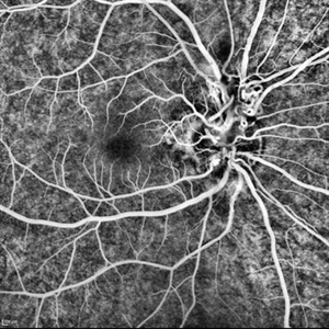 Peripapillary Vascular Loops
Jun 22 2018 by Hashim Ali Khan, OD, FAAO
FA of a 10-year-old boy with congenital peripapillary vascular loop.
Imaging device: Heidelberg Spectralis
Condition/keywords: congenital prepapillary vascular loop, pediatic retina
-
 Preretinal Vascular Loop
Feb 20 2015 by H. Michael Lambert, MD
Vascular loop on the optic nerve. 29-year-old black male, else normal eye exam. Father, sister and niece also had vascular abnormalities.
Condition/keywords: dominantly inherited, optic nerve, vascular loop
-
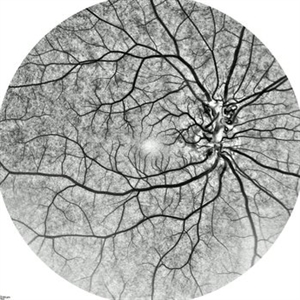 Congenital Peripapillary Vascular Loops
Jun 22 2018 by Hashim Ali Khan, OD, FAAO
Inverted FA of a 10-year-old boy with congenital peripapillary vascular loop.
Imaging device: Spectraliz
Condition/keywords: congenital prepapillary vascular loop, pediatic retina
-
 Vascular Loop
Feb 17 2014 by Howard Schatz, MD
79-year-old black female. Vascular loop. 20/64 OU.
Condition/keywords: vascular loop
-
 HTN Retinopathy with Pre-Papillary Vascular Loop OS
Jun 4 2018 by Hosam Attia, MD
Color fundus photograph of 53-year-old, African American male with history of diabetes, hypertension, depicting chronic hypertensive retinopathy changes and unilateral pre-papillary vascular loop OS.
Imaging device: Optos California
Condition/keywords: congenital prepapillary vascular anomaly, congenital prepapillary vascular loop, prepapillary vascular loop
-
 Autosomal Dominant Preretinal Vascular Loops
Jan 5 2015 by H. Michael Lambert, MD
Fluorescein angiogram showing corkscrew loop on right optic nerve.
Condition/keywords: vascular loop
-
 HTN Retinopathy with Pre-Papillary Vascular Loop OS
Jun 4 2018 by Hosam Attia, MD
Close-up color fundus photograph of 53-year-old, African American male with history of diabetes, hypertension, depicting chronic hypertensive retinopathy changes and unilateral pre-papillary vascular loop OS.
Imaging device: Optos California
Condition/keywords: congenital prepapillary vascular anomaly, congenital prepapillary vascular loop, prepapillary vascular loop
 Loading…