Search results (10 results)
-
 Rubella retinopathy
Dec 19 2012 by Eric A. Postel, MD
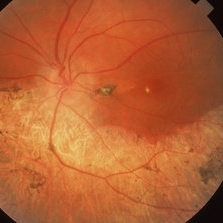
Color fundus photograph of a teenage girl with rubella retinopathy
Condition/keywords: rubella retinopathy, secondary pigmentary degeneration
-
 Pseudo Retinitis Pigmentosa
Apr 2 2019 by Gary R. Cook, MD, FACS
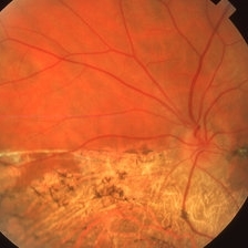
47-year-old white female with sectoral pigmentary degeneration inferiorly OS secondary to long-standing lattice-associated rhegmatogenous retinal detachment OS; V.A. = 20/70
Imaging device: Topcon VT-50
Condition/keywords: pseudo retinitis pigmentosa, secondary pigmentary degeneration
-
 Pseudo Retinitis Pigmentosa
Apr 2 2019 by Gary R. Cook, MD, FACS
47-year-old white female with a sectoral pigmentary degeneration inferiorly OS secondary to a long-standing, lattice-associated rhegmatogenous retinal detachment; V.A. = 20/70
Imaging device: Topcon VT-50
Condition/keywords: pseudo retinitis pigmentosa, secondary pigmentary degeneration
-
 Mellaril Toxicity
Apr 2 2019 by Gary R. Cook, MD, FACS
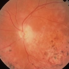
White male with a pigmentary retinopathy OS secondary to long-term treatment with Mellaril for psychosis.
Imaging device: Topcon VT-50
Condition/keywords: bilateral pigmentary retinopathy, mellaril toxicity, secondary pigmentary degeneration
-
 Mellaril Toxicity
Apr 2 2019 by Gary R. Cook, MD, FACS
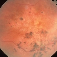
White male with a pigmentary retinopathy OD secondary to long-term treatment with Mellaril for psychosis.
Imaging device: Topcon VT-50
Condition/keywords: bilateral pigmentary retinopathy, mellaril toxicity, secondary pigmentary degeneration
-
 Elmiron Toxicity
Jan 15 2025 by Virginia Gebhart
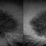
54 year old female with pigmentary degeneration secondary to Elmiron. Stippled RPE maculopathy has lightly progressed with stable vision compared to previous visits. BCVA 20/200 OU. Pt reports taking Elmiron from 2010 to 2019.
Photographer: Virginia Gebhart
Imaging device: Optos California
Condition/keywords: autofluorescence imaging, Maculopathy, secondary pigmentary degeneration
-
 Siderosis
Apr 2 2019 by Gary R. Cook, MD, FACS
27-year-old white male demonstrating pigment changes OS secondary to siderosis; V.A. = 20/30
Condition/keywords: secondary pigmentary degeneration, siderosis
-
 Pigmentary Degeneration of Retina (Secondary to Elmiron)
Nov 27 2024 by Virginia Gebhart
77 year old female with advanced geographic atrophy after years of Elmiron use (stopped in 2018). Serial exams show continued progression of GA. Central vision limited, vision remains stable and patient does not report noticing any changes.
Photographer: Virginia Gebhart, Retina Consultants of Carolina
Imaging device: Optos California
Condition/keywords: geographic atrophy, secondary pigmentary degeneration, toxic maculopathy
-
 Secondary Pigmentary Degeneration of Retina
Jul 18 2025 by Kimberly Wakester
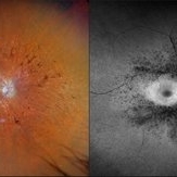
Optomap RGB and AF of an 63-year-old man with secondary pigmentary degeneration of the retina. Patient's Spark genetic testing revealed heterozygous mutations of unknown significance in LRP5, COL18A1, CPLANE1, SLC24A1 and VCAN. Clinical findings most consistent with Wagner's Syndrome (VCAN mutation, autosomal dominant). Will continue follow up care every 6 months with dilated exam and repeat OCT and Optos imaging.
Photographer: Kimberly Wakester, COA, OCT-C
Imaging device: Optos California
Condition/keywords: secondary pigmentary degeneration, Wagner's Syndrome
-
 Secondary Pigmentary Degeneration of Retina
Jul 18 2025 by Kimberly Wakester
Optomap RGB and AF of an 63-year-old man with secondary pigmentary degeneration of the retina. Patient's Spark genetic testing revealed heterozygous mutations of unknown significance in LRP5, COL18A1, CPLANE1, SLC24A1 and VCAN. Clinical findings most consistent with Wagner's Syndrome (VCAN mutation, autosomal dominant). Will continue follow up care every 6 months with dilated exam and repeat OCT and Optos imaging .
Photographer: Kimberly Wakester, COA, OCT-C
Imaging device: Optos California
Condition/keywords: secondary pigmentary degeneration, Wagner disease
 Loading…