Search results (80 results)
-
 Stage 5 Retinopathy of Prematurity (ROP)
Oct 9 2012 by Audina M. Berrocal, MD FASRS
Advanced APROP with Stage 5 and vascularly active.
Photographer: Ditte Hess CRA, BPEI
Imaging device: RETCAM
Condition/keywords: retinopathy of prematurity (ROP), stage 5
-
 Rhegmatogenous Retinal Detachment in Retinopathy of Prematurity
Oct 9 2012 by Audina M. Berrocal, MD FASRS
45-week-old ex-premature 24-week child who had a rhegmatogenous detachment after laser
Photographer: Ditte Hess CRA, BPEI
Imaging device: Ret Cam
Condition/keywords: laser, retinopathy of prematurity (ROP)
-
 Aggressive Posterior Retinopathy of Prematurity with Macular Hemorrhage
Oct 9 2012 by Audina M. Berrocal, MD FASRS
APROP with multiple pre-retinal hemorrhages
Photographer: Ditte Hess CRA, BPEI
Imaging device: RETCAM
Condition/keywords: macular hemorrhage, retinopathy of prematurity (ROP)
-
 Retinopathy of prematurity - dragged disc
Jan 11 2013 by Alex P. Hunyor, MD
Dragged disc due to retinopathy of prematurity.
Condition/keywords: dragged disc, retinopathy of prematurity (ROP)
-
 ROP Dragging
-
 Aggressive Posterior Retinopathy of Prematurity
Oct 9 2012 by Audina M. Berrocal, MD FASRS
Aggressive posterior Type 1 ROP with bleeding from regression of the posterior hyaloid artery
Photographer: Ditte Hess CRA, BPEI
Imaging device: RETCAM
Condition/keywords: retinopathy of prematurity (ROP)
-
 ROP Leukocoria
Oct 19 2012 by Larry Halperin, MD
ROP leukocoria
Condition/keywords: leukocoria, retinopathy of prematurity (ROP)
-
 Skip Area in Retinopathy of Pretmaturity
Oct 9 2012 by Audina M. Berrocal, MD FASRS
Skip area in ROP after laser treatment.
Photographer: Ditte Hess CRA, BPEI
Imaging device: RETCAM
Condition/keywords: laser, retinopathy of prematurity (ROP)
-
 Stage IVB
Oct 9 2012 by Audina M. Berrocal, MD FASRS
Progression of ROP to Stage IVB despite laser treatment.
Photographer: Ditte Hess CRA, BPEI
Imaging device: RetCam Digital Imaging
Condition/keywords: retinopathy of prematurity (ROP)
-
 Aggressive Posterior Retinopathy of Prematurity with Macular Hemorrhage
Oct 9 2012 by Audina M. Berrocal, MD FASRS
Aggressive posterior Type 1 ROP
Photographer: Ditte Hess CRA, BPEI
Imaging device: RETCAM
Condition/keywords: aggressive posterior retinopathy of prematurity (APROP), macular hemorrhage, retinopathy of prematurity (ROP)
-
 Retinopathy of Prematurity
Oct 19 2012 by Larry Halperin, MD
Retinopathy of prematurity
Condition/keywords: retinopathy of prematurity (ROP)
-
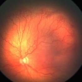
 Retinopathy of Prematurity Stage 3
Oct 23 2012 by Larry Halperin, MD
Retinopathy of prematurity, stage 3
Condition/keywords: retinopathy of prematurity (ROP), retinopathy of prematurity stage 3
-
 Latrogenic Oxygen Induce Retinopathy in a Premature Baby
Mar 13 2013 by Maria Ana Martinez-Castellanos, MD
Angiogram taken on a 42 weeks corrected age baby born at 34 weeks. The baby developed lung disease and received oxygen 100% for 4 weeks.
Photographer: Maria A. Martinez-Castellanos. Asociacion para Evitar la Ceguera en Mexico
Imaging device: RetCam II
Condition/keywords: retinopathy of prematurity (ROP)
-
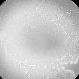
 Vascular loops in retinopathy of prematurity
Nov 3 2013 by Maria Ana Martinez-Castellanos, MD
Angiography of a baby with ROP treated with intravitreal anti-angiogenic therapy 1 week prior to the time this photo was taken. We can see the active vascular remodeling, vascular loops, the place where the demarcation line was at the time of the diagnosis and the growth of new vessels into the avascular zone, the leakage corresponds to immature vessels not fully covered by mural cells and not due to an inflammatory reaction.
Photographer: Maria A. Martinez-Castellanos. Asociacion para Evitar la Ceguera en Mexico
Imaging device: RetCam II
Condition/keywords: anti-VEGF, retinopathy of prematurity (ROP)
-
 Stage 1 ROP
Jun 4 2020 by Audina M. Berrocal, MD FASRS
Color photograph of the right eye in a premature baby. There is no plus disease, or evidence of Stage 1 in Zone 2 ROP. Stage 1 is defined as a line that separates vascularized retina to avascular retina.
Condition/keywords: retinopathy of prematurity (ROP), retinopathy of prematurity stage 1
-
 Retinopathy of Prematurity Under-Treated with Laser
Nov 8 2013 by Maria Ana Martinez-Castellanos, MD
Patient referred for a 2nd opinion after photocoagulation for ROP treatment. We can see under-treated avascular areas with no neovascular activity or elevated ridge
Photographer: Maria A. Martinez-Castellanos. Asociacion para Evitar la Ceguera en Mexico
Imaging device: RetCam II
Condition/keywords: laser photocoagulation, laser treatment, retinopathy of prematurity (ROP)
-
 Iridocorneal Angle
Sep 13 2013 by Maria Ana Martinez-Castellanos, MD
Angle OCT of a right eye of a 4 months old male patient with retinopathy of prematurity stage 5.
Photographer: Maria A. Martinez-Castellanos. Asociación para Evitar la Ceguera en Mexico
Condition/keywords: angle closure, retinopathy of prematurity (ROP), retinopathy of prematurity, stage 5
-
 Retinopathy of Prematurity Stage 4a
Sep 7 2013 by Maria Ana Martinez-Castellanos, MD
Retinopathy of prematurity stage 4a.
Photographer: Maria A. Martinez-Castellanos. Asociacion para Evitar la Ceguera en Mexico
Imaging device: RetCam II
Condition/keywords: retinopathy of prematurity (ROP), retinopathy of prematurity stage 4a
-
 Lobular Choroidal Filling in ROP Stage 1
Nov 3 2013 by Maria Ana Martinez-Castellanos, MD
Lobular choroidal filling in a baby with stage 1 ROP, the macular area has a different filling pattern that the rest of the retina.
Photographer: Maria A. Martinez-Castellanos. Asociacion para Evitar la Ceguera en Mexico
Imaging device: RetCam II
Condition/keywords: choriocapillaris, retinopathy of prematurity (ROP)
-
 Aggressive Posterior Retinopathy of Prematurity
May 8 2017 by Juan Romo-Aguas
Fundus photograph of an 1 month and 21 days female with bilateral aggresive posterior retinopathy of prematurity.
Photographer: Juan C. Romo-Aguas, Asociación Para Evitar la Ceguera en México
Imaging device: Optos Daytona Ultra-widefield Retinal Imaging
Condition/keywords: aggressive posterior retinopathy of prematurity (APROP), retinopathy of prematurity (ROP)
-
 Retinopathy of Prematurity Stage 2
Sep 7 2013 by Maria Ana Martinez-Castellanos, MD
Fluorescein angiography of a patient with ROP stage 2.
Photographer: Maria A. Martinez-Castellanos. Asociacion para Evitar la Ceguera en Mexico
Imaging device: RetCam II
Condition/keywords: retinopathy of prematurity (ROP)
-
 Screening Retinopathy of Prematurity with an Iphone 5
Dec 30 2013 by Maria Ana Martinez-Castellanos, MD
Telemedicine with smartphone.
Photographer: Maria A. Martinez-Castellanos. Asociacion para Evitar la Ceguera en Mexico
Imaging device: Smartphone with a 40 diopters lens
Condition/keywords: retinopathy of prematurity (ROP), retinopathy of prematurity stage2, smartphone fundus photography, telemedicine, video
-
 Fibrotic Tractional Membrane in ROP Stage 5
Nov 7 2013 by Maria Ana Martinez-Castellanos, MD
Stage 5 retinopathy of prematurity in a 6 month old baby.
Photographer: Maria A. Martinez-Castellanos. Asociacion para Evitar la Ceguera en Mexico
Imaging device: RetCam II
Condition/keywords: fibrous proliferation, fibrovascular proliferation, retinopathy of prematurity (ROP)
-
 Retinopathy of Prematurity in Zone 1
Feb 11 2015 by Darrell E. Baskin, MD
Retcam fundus photograph of a former 24-weeker (now 34 weeks), 740-gram birth weight showing retinopathy of prematurity in Zone 1 with pre-plus to plus disease (depending on examiner).
Photographer: Darrell Baskin, Wilford Hall, Lackland Air Force Base, Texas
Imaging device: Retcam 3
Condition/keywords: plus disease, retinopathy of prematurity (ROP)
-
---thumb.jpg/image-square;max$300,300.ImageHandler) Retinopathy of Prematurity
Retinopathy of Prematurity
Feb 5 2014 by Maurice F. Rabb
Baby born at 26 weeks gestation. The right eye reveals a large arteriole venous shunt in the peripheral retina. The venules leading away from the shunt appear to be larger than normal. Superimposed on the venules and arterioles are a number of small, round, reddish-pink 'bulbs' that are preretinal. Another interesting finding is the presence of a 'bulb' located on the disc. Angiography demonstrates that the lesions fluoresces vividly along with three other lesions, located 1 to 2 disc diameters from the disc. There was a vitreous hemorrhage in the left eye.
Condition/keywords: retinopathy of prematurity (ROP)
 Loading…