Search results (6 results)
-
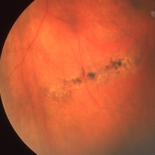
 Supero-Temporal Bullous Retinal Detachment With Macular Splitting
Sep 15 2017 by Somnath Chakraborty, MD
Montage fundus photo of a 56-year-old female with a bullous rhegmatogenous retinal detachment in the supero-temporal quadrant, secondary to a large horse shoe tear at 10 o' clock hour. She also has a large, pigmented lattice extending from 4 to 6 o' o' clock hours.
Photographer: Saptarshi Mehta, Retina Institute of Bengal
Condition/keywords: macular splitting, pigmented lattice lesion, retinal tear
-
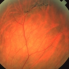
 Lattice Lesion
Nov 9 2012 by Norman Byer
This lattice lesion in a 70-year-old woman is almost entirely pigmented but several white lines can be faintly seen.
Condition/keywords: lattice degeneration, lattice lesion, pigmented lattice lesion, white lattice lines
-
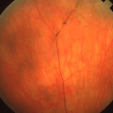
 Lattice Lesion
Nov 9 2012 by Norman Byer
This lattice lesion in a 44-year-old woman shows combined features of pigmentation, white lines, yellow dots and a round hole with a tiny zone of adjacent detachment. There are three such holes in this eye and they have not changed or been treated for eight years.
Condition/keywords: adjacent detachment, atrophic retinal hole, lattice degeneration, lattice lesion, pigmented lattice lesion, round hole, white lattice lines, yellow dots
-
 Pigmented Lattice Retinal Degeneration
Mar 27 2019 by Gary R. Cook, MD, FACS
54-year-old white female with pigmented lattice retinal lesion; V.A.= 20/20.
Imaging device: Topcon VT-50
Condition/keywords: lattice degeneration, pigmented lattice lesion
-
 Pigmented Paravascular Lattice
Mar 27 2019 by Gary R. Cook, MD, FACS
61-year-old white male with pigmented paravascular superiorly; V.A.= 20/25.
Imaging device: Topcon VT-50
Condition/keywords: lattice degeneration, pigmented lattice lesion
-
 Pigmented Paravascular Lattice
Mar 27 2019 by Gary R. Cook, MD, FACS
White female with peripheral lattice degeneration and some paravascular pigmentation.
Imaging device: Topcon VT-50
Condition/keywords: lattice degeneration, pigmented lattice lesion
 Loading…