Search results (79 results)
-
 Retinal Pigment Changes After Blunt Ocular Trauma
Jun 27 2016 by Rita Couceiro, MD, MS
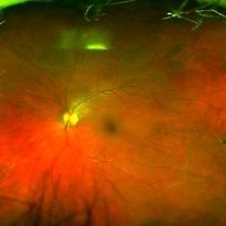
A 19-year-old man suffered blunt trauma of the left eye with a ball during soccer practice. At day 3 after trauma (upper pictures) the retinal area superior to the fovea looked pale and visual acuity was reduced to 20/32. This area revealed hypersignaling of retinal layers on OCT and the foveal area showed a localized disruption of retinal layers above the RPE. At day 30 (lower pictures) the retinal area of pallor showed pigmentary changes and OCT revealed atrophy of the external retinal layers. However the localized subfoveal retinal disruption was improved and only a slight disruption was seen on OCT at the ellipsoid level. Visual acuity of the left eye was restored to 20/20 although a scotoma remained.
Photographer: Rita Couceiro, Serviço de Oftalmologia do Hospital de Santa Maria, Lisboa, Portugal
Condition/keywords: blunt trauma, commotio retinae, pigment changes
-
 Optic Nerve Pit
Aug 30 2012 by Raj K. Maturi, MD
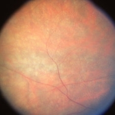
congenital optic nerve pit with chronic pigment changes in macula due to detachment
Photographer: Tom Steele, CRA, Midwest Eye Institute
Imaging device: Topcon Ex
Condition/keywords: optic nerve pit
-
---thumb.JPG/image-square;max$300,300.ImageHandler) Measles Retinopathy
Measles Retinopathy
Jun 29 2013 by Jason S. Calhoun
52-year-old female wanted second opinion on retinal eval. Patient had PK and PDT in the left eye. VA was 20/30, right eye was count fingers. Pinhole was NI, both eyes. Patient's mother had measles. Notice pigment changes in both the color and FAF photo.
Photographer: Jason S. Calhoun, Mayo Clinic Jacksonville, Florida
Imaging device: TOPCON TRC 50-EX
Condition/keywords: measles retinopathy
-
 Peripheral, linear, pigment changes
Apr 4 2013 by Jerald A. Bovino, MD
Peripheral, linear, pigment changes
Condition/keywords: pigment changes
-
---thumb.JPG/image-square;max$300,300.ImageHandler) Dry Macular Degeneration With Hemorrhage
Dry Macular Degeneration With Hemorrhage
Jul 13 2013 by Jason S. Calhoun
Pigment changes in the macula with hemorrhages present temporally.
Photographer: Jason S. Calhoun, Department of Ophthalmology, Mayo Clinic Jacksonville, Florida
Imaging device: TOPCON TRC 50-EX
Condition/keywords: dry age-related macular degeneration (dry AMD)
-
 Central atrophic pigment changes
Apr 4 2013 by Jerald A. Bovino, MD
No history, seems to have flecks, probably hereditary macular degeneration
Condition/keywords: pigment changes
-
 Choroideremia With Periperal Pigment Changes, Drusenoid Flecks, Patchy Atrophy
Aug 1 2013 by From the Collections of Thomas M. Aaberg, MD and Thomas M. Aaberg Jr., MD
Choroideremia with periperal pigment changes, drusenoid flecks, patchy atrophy.
Condition/keywords: atrophy, choroideremia, drusenoid flecks
-
 Measles Retinopathy
Jun 29 2013 by Jason S. Calhoun
52-year-old female wanted second opinion on retinal eval. Patient had PK and PDT in the left eye. VA was 20/30, right eye was count fingers. Pinhole was NI, both eyes. Patient's mother had measles. Notice pigment changes in both the color and FAF photo.
Photographer: Jason S. Calhoun, Mayo Clinic Jacksonville, Florida
Imaging device: TOPCON TRC 50-EX
Condition/keywords: measles retinopathy
-
 Drusen
Apr 4 2013 by Jerald A. Bovino, MD
No history, pigment changes, probably drusen
Condition/keywords: pigment changes
-
 Widespread pigment changes
Apr 4 2013 by Jerald A. Bovino, MD
No history, the ON is pink and vessels look normal
Condition/keywords: pigment changes
-
 Pigment Dispersion Syndrome
May 25 2016 by M. Reza Razeghinejad
Fundus photograph of a 57-year-old woman with pigment dispersion syndrome and retinal perivascular pigmentation
Condition/keywords: pigment changes
-
 Choroideremia With Periperal Pigment Changes, Drusenoid Flecks, Patchy Atrophy
Aug 1 2013 by From the Collections of Thomas M. Aaberg, MD and Thomas M. Aaberg Jr., MD
Choroideremia with periperal pigment changes, drusenoid flecks, patchy atrophy.
Condition/keywords: atrophy, choroideremia, drusenoid flecks
-
 Widespread pigment changes
Apr 4 2013 by Jerald A. Bovino, MD
the ON is pink and vessels look normal
Condition/keywords: pigment changes
-
 Choroideremia With Periperal Pigment Changes, Drusenoid Flecks, Patchy Atrophy
Aug 1 2013 by From the Collections of Thomas M. Aaberg, MD and Thomas M. Aaberg Jr., MD
Choroideremia with periperal pigment changes, drusenoid flecks, patchy atrophy.
Condition/keywords: atrophy, choroideremia, drusenoid flecks
-
 central pigment epithelial changes
Apr 4 2013 by Jerald A. Bovino, MD
central pigment epithelial changes
Condition/keywords: epithelial changes, pigment changes
-
 Central Pigment Changes
-
 Choroideremia With Periperal Pigment Changes, Drusenoid Flecks, Patchy Atrophy
Aug 1 2013 by From the Collections of Thomas M. Aaberg, MD and Thomas M. Aaberg Jr., MD
Choroideremia with periperal pigment changes, drusenoid flecks, patchy atrophy.
Condition/keywords: atrophy, choroideremia, drusenoid flecks
-
 Choroideremia Carrier
Aug 1 2013 by From the Collections of Thomas M. Aaberg, MD and Thomas M. Aaberg Jr., MD
FA of choroideremia carrier with diffuse mottled fluorescence from pigment changes
Condition/keywords: choroideremia, pigment changes
-
 Pigmentary Retinal Dystrophy
Mar 29 2019 by Jessica Norkus
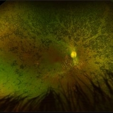
Optos ultra wide field image of 41-year-old male patient with pigmentary retinal dystrophy. Atypical findings due to unilateral presentation. Patient has been experiencing symptoms for 15 years, notes significant nyctalopia.
Photographer: Jessica Norkus
Imaging device: Optos Ultra Wide Field Camera
Condition/keywords: abnormal fundus, bone spicule, color fundus photograph, color photo, fundus photograph, Optos, peripheral bone spicules, pigment changes, ultra-wide field imaging, unilateral blindness
-
 Choroideremia With Periperal Pigment Changes, Drusenoid Flecks, Patchy Atrophy
Aug 1 2013 by From the Collections of Thomas M. Aaberg, MD and Thomas M. Aaberg Jr., MD
Choroideremia with periperal pigment changes, drusenoid flecks, patchy atrophy.
Condition/keywords: atrophy, choroideremia, drusenoid flecks
-
 central pigment epithelial changes
-
 Central Pigment Changes
Jul 11 2013 by Jerald A. Bovino, MD
No history, upside down and high contrast.
Condition/keywords: pigment changes
 Loading…