Search results (12 results)
-
 Snail Track Peripheral Retinal Degeneration
Apr 29 2022 by Otakar Dušek, M.D. Ph.D.
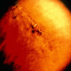
Colour fundus photograph of 22-year-old woman with incidentally found snail track retinal degeneration in the superior temporal periphery of the retina of the right eye.
Photographer: Otakar Dušek, Charles University, Prague
Imaging device: Zeiss Clarus
Condition/keywords: peripheral retinal degeneration
-
 Pigmented Peripheral Retinal Degeneration
Jun 27 2013 by Jason S. Calhoun
42-year-old male came in for routine eye exam and to follow up on peripheral retinal degeneration in both eyes. VA is 20/20, right eye and 20/25, left eye. Patient is asymptomatic with no visual complaints.
Photographer: Jason S. Calhoun, Mayo Clinic Jacksonville, Florida
Imaging device: TOPCON TRC 50-EX
Condition/keywords: peripheral retinal degeneration
-
---thumb.JPG/image-square;max$300,300.ImageHandler) Peripheral Retinal Degeneration
Peripheral Retinal Degeneration
Jul 8 2013 by Jason S. Calhoun
Patient comes in with double vision. VA was 20/20 in both eyes. Fundus exam shows retinal degenerative changes in both eyes. Offer to correct double vision with temporary Fresnel prism.
Photographer: Jason S. Calhoun, Department of Ophthalmology, Mayo Clinic Jacksonville, Florida
Condition/keywords: peripheral retinal degeneration
-
 Lattice Degeneration
May 2 2013 by Henry J. Kaplan, MD
Pigmented lattice degeneration with lattice "wicker" caused by sclerotic blood vessels.
Condition/keywords: lattice degeneration, peripheral retinal degeneration
-
 ARMD With Geographic Atrophy, Peripheral Degeneration
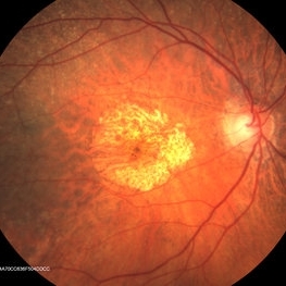
Dec 6 2013 by James B. Soque, CRA, OCT-C, COA, FOPS
92-year-old white female with exudative macular degeneration, geographic atrophy, and peripheral retinal degeneration.
Photographer: James Soque, CRA COA, Island Retina, Shirley, New York
Imaging device: Topcon TRC 50DX with OIS 10.6.45
Condition/keywords: geographic atrophy, macular degeneration, retinal degeneration
-
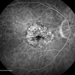 ARMD With Geographic Atrophy , Peripheral Degeneration, FA
Dec 6 2013 by James B. Soque, CRA, OCT-C, COA, FOPS
FA right eye early phase, of a 92-year-old white female with exudative macular degeneration, geographic atrophy, and peripheral retinal degeneration.
Photographer: James Soque CRA COA, Island Retina, Shirley, New York
Imaging device: Topcon TRC 50DX with OIS 10.6.45
Condition/keywords: geographic atrophy
-
 Peripheral Retinal Degeneration
Jul 8 2013 by Jason S. Calhoun
Patient in with double vision. VA was 20/20 in both eyes. Fundus exam shows retinal degenerative changes in both eyes. Offer to correct double vision with temporary Fresnel prism.
Photographer: Jason S. Calhoun, Department of Ophthalmology, Mayo Clinic Jacksonville, Florida
Condition/keywords: peripheral retinal degeneration
-
 ARMD With Geographic Atrophy, Peripheral Degeneration
Dec 6 2013 by James B. Soque, CRA, OCT-C, COA, FOPS
92-year-old white female with exudative macular degeneration, geographic atrophy, and peripheral retinal degeneration.
Photographer: James Soque, CRA COA, Island Retina, Shirley, New York
Imaging device: Topcon TRC 50DX with OIS 10.6.45
Condition/keywords: fundus photograph, geographic atrophy
-
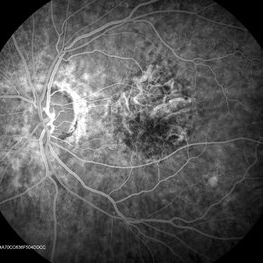 ARMD With Geographic Atrophy, Peripheral Degeneration, FA
Dec 6 2013 by James B. Soque, CRA, OCT-C, COA, FOPS
FA left eye Early Phase, of a 92-year-old white female with exudative macular degeneration, geographic atrophy, and peripheral retinal degeneration.
Photographer: James Soque CRA COA, Island Retina, Shirley, New York
Imaging device: Topcon TRC 50DX with OIS 10.6.45
Condition/keywords: geographic atrophy
-
 White Without Pressure and Peripheral Retinoschisis
Dec 29 2022 by Gulnara Islamova
Fundus Photograph and OCT scan of an 18 year-old male with peripheral retinoschisis combined with WWOP lessions .Vitreoretinal traction is not visualized
Photographer: Gulnara Islamova, CENTER ZRENIYA Medical Clinic, LLC, Chelyabinsk, Russian Federation
Imaging device: Optovue XR Avanti
Condition/keywords: peripheral retinal degeneration
-
 Peripheral retinal degenerations
Jan 29 2024 by Anupama Kiran Kumar
Fundus photo of a young man who underwent barrage laser after he presented to the clinic with floaters and was diagnosed to have lattices with horse shoe tears and retinal holes.
Photographer: Dr Anupama Kiran Kumar DNB FVR , Narayana Nethralaya Bangalore
Imaging device: Mirante SLO/OCT (Nidek Co., Gamagori, Japan)
Condition/keywords: lattice degeneration, peripheral retinal degeneration
-
 Peripheral Retinal Degeneration (L-ORD)
Apr 17 2024 by Virginia Gebhart
92 year old female with bilateral patchy, sharply demarcated circular areas of chorioretinal atrophy with hyperpigmented margins in the mid to far periphery. Labs showed normal plasma ornithine levels ruling out generalized gyrate atrophy. Also intermediate uveitis and CMD/CME. FTA-ABS, Quant gold, and HLA-A29 labs all negative.
Photographer: Virginia Gebhart
Imaging device: Optos California
Condition/keywords: cystoid macular degeneration, cystoid macular edema (CME), FA, Fluorescein angiography, peripheral retinal degeneration
 Loading…