Search results (54 results)
-
 Acquired Optic Pit Maculopathy
Aug 20 2014 by Andree Henaine-Berra, MD
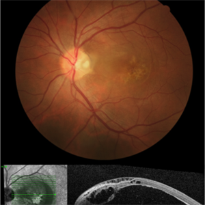
Optical coherence tomography of the left eye of a 60-year-old man with an acquired optic pit maculopathy and glaucoma. The image shows an enlarged optic disc cup and a macular serous detachment.
Photographer: Andree Henaine-Berra. Asociacion Para Evitar la Ceguera en Mexico. Mexico City.
Imaging device: Heidelberg Spectralis
Condition/keywords: glaucoma, maculopathy, optic pit
-
 Optic Pit FA
Jul 4 2012 by John T. Thompson, MD
Hyperfluorescence in optic pit due to fluorescein leakage
Imaging device: Zeiss FF4
Condition/keywords: fluorescein leakage, optic disc pit
-
 Optic Pit
Oct 12 2012 by Gregg T. Kokame, MD, MMM, FASRS
Optic pit
Photographer: Jaclyn Pisano, Retina Consultants of Hawaii
Imaging device: Zeiss FF-450 plus
Condition/keywords: congenital optic nerve pit
-
 Optic Pit With Old Resolved Subretinal Fluid
Feb 20 2013 by From the Collections of Thomas M. Aaberg, MD and Thomas M. Aaberg Jr., MD
20/200.
Condition/keywords: optic disc pit, resolved subretinal fluid
-
 Optic Pit; two in one nerve
Feb 19 2013 by From the Collections of Thomas M. Aaberg, MD and Thomas M. Aaberg Jr., MD
Color photo, 20/20; left of a stereo pair.
Condition/keywords: color photo, stereo pair
-
 Optic Pit; two in one nerve
Feb 19 2013 by From the Collections of Thomas M. Aaberg, MD and Thomas M. Aaberg Jr., MD
Color photo, 20/20; left of a stereo pair.
Condition/keywords: color photo, stereo pair
-
---thumb.JPG/image-square;max$300,300.ImageHandler) Optic Pit Maculopathy - Fundus Photograph
Optic Pit Maculopathy - Fundus Photograph
Oct 14 2013 by Cagri G Besirli, MD, PhD, FASRS
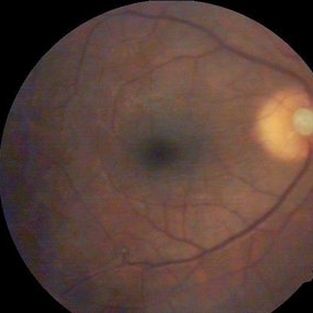
10-year-old girl with congenital optic pit and recent vision loss secondary to optic pit maculopathy.
Imaging device: Optos
Condition/keywords: fundus photograph, maculopathy
-
 Optic Pit With Subretinal Fluid and Macular Cyst
Feb 20 2013 by From the Collections of Thomas M. Aaberg, MD and Thomas M. Aaberg Jr., MD
No history.
Condition/keywords: optic disc pit, subretinal fluid
-
 Optic Pit Maculopathy
Jan 8 2017 by Mario Canastro
Fundus photograph and spectral-domain OCT of an 43-year-old male with left eye optic pit maculopathy.
Photographer: Mário Canastro, MD, MSc - Serviço de Oftalmologia do Hospital de Santa Maria, Lisboa, Portugal
Imaging device: Fundus camera and Heidelberg Spectralis OCT
Condition/keywords: maculopathy, optic pit
-
 Optic Pit; two in one nerve
Feb 19 2013 by From the Collections of Thomas M. Aaberg, MD and Thomas M. Aaberg Jr., MD
Color photo, 20/15; fellow eye.
Condition/keywords: color photo, fellow eye
-
---thumb.JPG/image-square;max$300,300.ImageHandler) Optic Pit Maculopathy - Fundus Autofluorescence
Optic Pit Maculopathy - Fundus Autofluorescence
Oct 14 2013 by Cagri G Besirli, MD, PhD, FASRS
10-year-old girl with congenital optic pit and recent vision loss secondary to optic pit maculopathy.
Imaging device: Optos
Condition/keywords: fundus autofluorescence (FAF), maculopathy
-
 Optic pit
May 2 2013 by Henry J. Kaplan, MD
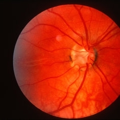
Optic pit in the inferotemporal part of the optic disc.
Condition/keywords: optic disc pit
-
 Optic Pit Red Free Photo
Jan 9 2014 by Susanna S. Park, MD, PhD
Red-free fundus photograph of a young man with recent vision loss from maculopathy associated with optic disc pit. Macular schisis and detachment with outer lamellar hole was noted preoperatively
Photographer: Ellen Redenbo, University of California Davis
Imaging device: Topcon
Condition/keywords: lamellar macular hole, macular schisis, optic disc pit, subretinal fluid
-
 Acquired Optic Pit Maculopathy
Aug 20 2014 by Andree Henaine-Berra, MD
Optical coherence tomography of the left eye of a 60-year-old man with an acquired optic pit maculopathy and glaucoma. The image shows subretinal fluid extending to the optic nerve and schisis of the outer retinal layers.
Photographer: Andree Henaine-Berra. Asociacion Para Evitar la Ceguera en Mexico. Mexico City.
Imaging device: Heidelberg Spectralis
Condition/keywords: glaucoma, maculopathy, optic pit
-
---thumb.jpg/image-square;max$300,300.ImageHandler) OCT Optic Pit Maculopathy Post-op
OCT Optic Pit Maculopathy Post-op
Jan 10 2014 by Susanna S. Park, MD, PhD
OCT image taken 1 year after vitrectomy with gas tamponade for macular schisis and detachment and outer lamellar hole associated with optic pit shows normal macular morphology with only mild disruption of the foveal photoreceptor layer.
Photographer: Ellen Redenbo, University of California Davis Eye Center
Condition/keywords: macular schisis, maculopathy, optical coherence tomography (OCT)
-
 Acquired Optic Pit Maculopathy
Aug 20 2014 by Andree Henaine-Berra, MD
Fundus photograph of the left eye of a 60-year-old man with an acquired optic pit maculopathy and glaucoma. The image shows an enlarged optic disc cup and a macular serous detachment.
Photographer: Andree Henaine-Berra. Asociacion Para Evitar la Ceguera en Mexico. Mexico City.
Imaging device: Heidelberg Spectralis
Condition/keywords: glaucoma, maculopathy, optic pit
-
---thumb.jpg/image-square;max$300,300.ImageHandler) OCT Optic Pit Maculopathy Preop
OCT Optic Pit Maculopathy Preop
Jan 10 2014 by Susanna S. Park, MD, PhD
Cirrus OCT of a 25-year-old man presenting with recent vision loss from severe macular schisis, outer lamellar hole and foveal detachment associated with an optic pit
Photographer: Ellen Redenbo, University of California Davis
Imaging device: Cirrus OCT
Condition/keywords: macular schisis, optical coherence tomography (OCT), outer lamellar hole
-
 Optic Pit
Jul 13 2016 by PAVEL FLORES-MORENO
OCT of a 56-year-old male with 7 days of low visual acuity.
Photographer: Flores-Moreno Pavel
Condition/keywords: optic pit, serous retinal detachment
-
 Acquired Optic Pit Maculopathy
Aug 20 2014 by Andree Henaine-Berra, MD
Autofluorescence image of the left eye of a 60-year-old man with an acquired optic pit maculopathy and glaucoma.
Photographer: Andree Henaine-Berra. Asociacion Para Evitar la Ceguera en Mexico. Mexico City.
Imaging device: Heidelberg Spectralis
Condition/keywords: glaucoma, maculopathy, optic pit
-
 Vertical OCT Scan Through Right Optic Disc Pit
Jul 20 2019 by Arwa Azmeh, MD, PhD
Fundus photograph of 38-year-old healthy man with right optic disc pit, who recently noticed slightly blurred vision in right eye while closing the left eye. BCVA was 20/25 in OD and 20/20 in OS. IOP was 15mmHg OD and 14 mmHg OS. Right fundus exam showed small optic disc pit near the temporal rim of optic disc with abnormal reflex of nasal macula. Left fundus was normal. Late FA of right optic disc showed no leakage or staining of optic disc. Macular OCT showed normal foveal contour with no subretinal fluid or macular edema. There was significant reduction in RNFL thickness in the temporal sector in right eye. Coloboma is clearly seen on vertical OCT scan as well as horizontal scans through right optic pit.
Photographer: Ebtisam Aljbeili, Damascus university, Almouassat university hospital
Imaging device: Heidelberg Spectralis 2
Condition/keywords: optic pit, optical coherence tomography (OCT)
-
 Optic Pit Large Fundus Photo
Jan 9 2014 by Susanna S. Park, MD, PhD
Magnfied fundus photograph of the disc showing a large optic pit on the disc.
Photographer: Ellen Redeenbo, University of California Davis Eye Center
Imaging device: Topcon
Condition/keywords: optic pit
-
 Optic Pit 2
Oct 10 2013 by Roy Schwartz, MD
Late phase FA of a young patient with optic pit.
Photographer: galit yair pur
Condition/keywords: early phase
-
---thumb.JPG/image-square;max$300,300.ImageHandler) Optic Pit Maculopathy - SD-OCT
Optic Pit Maculopathy - SD-OCT
Oct 14 2013 by Cagri G Besirli, MD, PhD, FASRS
10-year-old girl with congenital optic pit and recent vision loss secondary to optic pit maculopathy.
Imaging device: Heidelberg Spectralis
Condition/keywords: maculopathy, optical coherence tomography (OCT)
-
 Horizontal OCT Scan Through Right Optic Pit
Jul 20 2019 by Arwa Azmeh, MD, PhD
Fundus photograph of 38-year-old healthy man with right optic disc pit, who recently noticed slightly blurred vision in right eye while closing the left eye. BCVA was 20/25 in OD and 20/20 in OS. IOP was 15mmHg OD and 14 mmHg OS. Right fundus exam showed small optic disc pit near the temporal rim of optic disc with abnormal reflex of nasal macula. Left fundus was normal. Late FA of right optic disc showed no leakage or staining of optic disc. Macular OCT showed normal foveal contour with no subretinal fluid or macular edema. There was significant reduction in RNFL thickness in the temporal sector in right eye. Coloboma is clearly seen on vertical OCT scan as well as horizontal scans through right optic pit.
Photographer: Ebtisam Aljbeili, Damascus university, Almouassat university hospital
Imaging device: Heidelberg Spectralis 2
Condition/keywords: optic pit, optical coherence tomography (OCT)
-
 Optic Pit
Oct 4 2014 by Mehul A Shah
A 40-year-old male presented for routine check up and this finding occurred.
Photographer: Drashti Netralaya,Dahod
Imaging device: Zeiss ff450
Condition/keywords: optic pit
 Loading…