Search results (130 results)
-
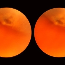 Enclosed Ora Bay On The Temporal Side
Nov 9 2012 by Norman Byer
This is another example of an enclosed ora bay on the temporal side. It is surrounded by normal retina and well separated from the ora serrata, which is toward the upper right just beyond the photograph. The yellow nubbin marks an abortive dentate process.
Condition/keywords: abortive dentate process, enclosed ora bay, normal eye, normal retina, ora serrata, temporal retina
-
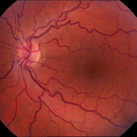
---thumb.jpg/image-square;max$300,300.ImageHandler) Normal Fundus Photo
Normal Fundus Photo
Feb 13 2013 by From the Collections of Thomas M. Aaberg, MD and Thomas M. Aaberg Jr., MD
Normal fundus photo.
Condition/keywords: fundus photograph, normal eye
-
 Scleral Indentation In A Normal Eye
Nov 9 2012 by Norman Byer
This shows the appearance of scleral indentation in a normal eye. Note the convex shadow which marks the posterior border of the indented area. It is caused in part by a small angle which separates the viewing axis from the illuminating axis thus allowing the observer to see slightly into the shadow beyond the illuminated crest of the indentation. It is also caused in part by viewing the pigment epithelial layer in a tangential manner. This shadow is of great diagnostic usefulness since it becomes a dark background against which many tiny retinal abnormalities can be seen beautifully by contrast. Two other particular advantages of scleral indentation will be demonstrated in the following photographs: First, the ability to see the extreme anterior part of the retina to the ora serrata and beyond, and second, the ability to examine any abnormality in multiple profiles depending on slight movements of the scleral depressor in various directions.
Condition/keywords: extreme anterior retina, posterior border, scleral indentation, shadow, tangential view of pigment epithelial layer
-
 Congénital Venous Tortuosity OS
Mar 13 2013 by Jose Dalma-Weiszhausz, MD
Young patient with routine normal eye exam.
Photographer: José Dalma, MD Dalma & Assoc., Mexico City
Imaging device: Topcon 50VT
Condition/keywords: congenital venous tortuosity
-
 Preretinal Vascular Loop
Feb 20 2015 by H. Michael Lambert, MD
Vascular loop on the optic nerve. 31-year-old black female, else normal eye exam. Father, brother and daughter also had vascular abnormalities.
Condition/keywords: dominantly inherited, optic nerve, vascular loop
-
 Normal Eye
Aug 27 2012 by Suber S. Huang, MD, MBA, FASRS
Photographer: Geoffrey Pankhurst, Case Western Reserve University/University Hospitals of Cleveland, Cleveland, OH
Imaging device: Topcon
Condition/keywords: normal eye
-
 Normal Eye
Aug 27 2012 by Suber S. Huang, MD, MBA, FASRS
Photographer: Geoffrey Pankhurst, Case Western Reserve University/University Hospitals of Cleveland, Cleveland, OH
Imaging device: Topcon
Condition/keywords: normal eye
-
 Normal Eye
Aug 27 2012 by Suber S. Huang, MD, MBA, FASRS
Photographer: Geoffrey Pankhurst, Case Western Reserve University/University Hospitals of Cleveland, Cleveland, OH
Imaging device: Topcon
Condition/keywords: normal eye
-
 Preretinal Vascular Loop
Feb 20 2015 by H. Michael Lambert, MD
Vascular loop on the optic nerve. 31-year-old black female, else normal eye exam. Father, brother and daughter also had vascular abnormalities.
Condition/keywords: dominantly inherited, optic nerve
-
 Preretinal Vascular Loop
Feb 20 2015 by H. Michael Lambert, MD
Vascular loop on the optic nerve. 29-year-old black male, else normal eye exam. Father, sister and niece also had vascular abnormalities.
Condition/keywords: dominantly inherited, optic nerve, vascular loop
-
 Normal Eye
Aug 27 2012 by Suber S. Huang, MD, MBA, FASRS
Photographer: Geoffrey Pankhurst, Case Western Reserve University/University Hospitals of Cleveland, Cleveland, OH
Imaging device: Topcon
Condition/keywords: normal eye
-
 Normal eye
-
 Normal Eye
Aug 27 2012 by Suber S. Huang, MD, MBA, FASRS
Photographer: Geoffrey Pankhurst, Case Western Reserve University/University Hospitals of Cleveland, Cleveland, OH
Imaging device: Topcon
Condition/keywords: normal eye
-
 Looks Pretty Normal
-
 Normal Eye
Aug 27 2012 by Suber S. Huang, MD, MBA, FASRS
Photographer: Geoffrey Pankhurst, Case Western Reserve University/University Hospitals of Cleveland, Cleveland, OH
Imaging device: Topcon
Condition/keywords: normal eye
-
 Normal
Jul 22 2013 by Howard Schatz, MD
24-year-old white male, right eye 20/25, left eye 20/20.
Condition/keywords: normal eye
-
 Normal
Jul 22 2013 by Howard Schatz, MD
Cilio ret. art.
Condition/keywords: cilioretinal artery occlusion, normal eye
-
 Preretinal Vascular Loop
Feb 20 2015 by H. Michael Lambert, MD
Vascular loop on the optic nerve. 31-year-old black female, else normal eye exam. Father, brother and daughter also had vascular abnormalities.
Condition/keywords: dominantly inherited, optic nerve, vascular loop
-
 Normal Fundus
Mar 26 2019 by Gary R. Cook, MD, FACS
Normal fundus photograph OS; VA= 20/20.
Imaging device: Topcon VT-50
Condition/keywords: normal eye
-
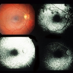
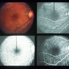
---thumb.jpg/image-square;max$300,300.ImageHandler) Fluoresecein Angiography
Fluoresecein Angiography
Feb 20 2013 by From the Collections of Thomas M. Aaberg, MD and Thomas M. Aaberg Jr., MD
Fluorescein angiography showing a normal venous phase flow shot.
Condition/keywords: normal eye, venous flow phase
-
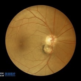 Double disc sign
Oct 13 2022 by Vaibhavi Noticewala, M S Ophthalmology, FVRS
Double disc sign Doubling of the optic disc is rare and can manifest as true or pseudo doubling. Duke-Elder describes duplication of the optic disc as a rare anomaly wherein two discs, each provided with retinal vessels are seen in an otherwise normal eye. Rare cases of true duplication of optic discs with separation of optic nerve into two or more strands have been reported, based either on incidental necropsy findings, demonstration of two optic foramina in the same orbit on x ray, or angioscotomas as indirect evidence of the existence of double optic nerves. Pseudo doubling of the optic discs caused by lesions such as optic disc coloboma, peripapillary chorioretinal coloboma, or inflammatory foci are more common. Our case had Ipsilateral isolated ectatic peripapillary chorioretinal coloboma simulating double optic discs.
Photographer: Priyal Mistry
Condition/keywords: Pseudoduplication of optic disc
-
 Normal
-
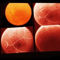
 Normal F/A
-
 Preretinal Vascular Loop
Feb 20 2015 by H. Michael Lambert, MD
29-year-old black male, else normal eye exam. Father, sister and niece also had vascular abnormalities.
Condition/keywords: dominantly inherited, optic nerve, vascular loop
-
 Partial Vitreous Separation in a High Myope With a Posterior Staphyloma
Dec 10 2012 by Yale L. Fisher, MD
This B-scan demonstrates a partial PVD. A posterior vitreous detachment (PVD) may occur in a normal aging eye or may be associated with pathology such as vitreous hemorrhage or inflammation. In a normal eye, as in this example, the PVD appears as a thin and smooth line (arrow) on B-scan. When the globe is moved voluntarily by the patient, real time echography demonstrates a quick jerky motion of the sheet-like echo with movements continuing after the globe movement has ceased. This is helpful in differentiating a PVD from a retinal detachment, which typically has a slower undulating pattern of motion. If there was presence of blood or inflammatory debris associated with the PVD, the echogenic line might appear thicker, especially in the most gravity dependent portions of the globe (i.e., posterior and inferior).
Condition/keywords: video
 Loading…