Search results (19 results)
-
 OCT Myopic Staphyloma With Schisis and ERM
Apr 24 2014 by Scott E. Pautler, MD
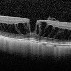
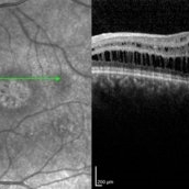
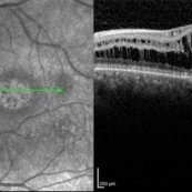
OCT of high myope with asymptomatic macular schisis.
Imaging device: Heidelberg Spectralis
Condition/keywords: foveal schisis, maculopathy, maculoschisis, optical coherence tomography (OCT), pathologic myopia, staphyloma
-
 Optic Disc Coloboma
Apr 25 2017 by Nimrod Dar
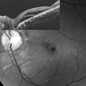
9 year-old patient, noticed a gradual deterioration in her visual acuity at her LE (6/15). On her examination, a double optic disc can be seen. OCT scan revealed an intra retinal fluid and macular schisis.
Photographer: Nimrod Dr, MD
Condition/keywords: coloboma of the optic nerve
-
 Myopic Macular Schisis with Lamellar Macular Hole
May 26 2014 by John T. Thompson, MD
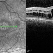
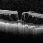
Spectral domain OCT of patient with high myopia and myopic macular schisis resulting in lamellar macular hole.
Condition/keywords: lamellar macular hole, myopic macular schisis
-
 Macular Schisis
Jan 23 2014 by Sharon Fekrat, MD FACS FASRS
SD-OCT image of macular schisis in the left eye of a 46-year-old female who is a -13D myope. Vision was 20/64.
Photographer: Duke Eye Imaging, Duke University Eye Center
Condition/keywords: macular schisis
-
 Optic Disc Pit Schisis RD
Apr 29 2013 by Michael Colucciello, MD, FASRS
Optic disc pit with peripapillary RD and macular schisis, fundus photograph and SD-OCT overlay.
Condition/keywords: optic disc pit
-
 Optic Pit Red Free Photo
Jan 9 2014 by Susanna S. Park, MD, PhD
Red-free fundus photograph of a young man with recent vision loss from maculopathy associated with optic disc pit. Macular schisis and detachment with outer lamellar hole was noted preoperatively
Photographer: Ellen Redenbo, University of California Davis
Imaging device: Topcon
Condition/keywords: lamellar macular hole, macular schisis, optic disc pit, subretinal fluid
-
---thumb.jpg/image-square;max$300,300.ImageHandler) OCT Optic Pit Maculopathy Post-op
OCT Optic Pit Maculopathy Post-op
Jan 10 2014 by Susanna S. Park, MD, PhD
OCT image taken 1 year after vitrectomy with gas tamponade for macular schisis and detachment and outer lamellar hole associated with optic pit shows normal macular morphology with only mild disruption of the foveal photoreceptor layer.
Photographer: Ellen Redenbo, University of California Davis Eye Center
Condition/keywords: macular schisis, maculopathy, optical coherence tomography (OCT)
-
 Juvenile Retinoschisis
Oct 10 2015 by Hamid Ahmadieh, MD
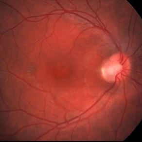
Color fundus photograph of the left eye of a 30-year-old man with juvenile retinoschisis and typical macular changes.
Photographer: Shabnam Pooreh, Negah Eye Center, Tehran , Iran
Condition/keywords: color fundus photograph, juvenile retinoschisis, macular schisis
-
 Macular Pseudo hole With Schisis
Aug 26 2018 by John T. Thompson, MD
Spectral domain OCT of macular pseudohole with epiretinal membrane and macular schisis. This would be classified as epiretinal membrane and foveoschisis in the international classification system.
Imaging device: Heidelberg Spectralis
Condition/keywords: epiretinal membrane (ERM)
-
---thumb.jpg/image-square;max$300,300.ImageHandler) OCT Optic Pit Maculopathy Preop
OCT Optic Pit Maculopathy Preop
Jan 10 2014 by Susanna S. Park, MD, PhD
Cirrus OCT of a 25-year-old man presenting with recent vision loss from severe macular schisis, outer lamellar hole and foveal detachment associated with an optic pit
Photographer: Ellen Redenbo, University of California Davis
Imaging device: Cirrus OCT
Condition/keywords: macular schisis, optical coherence tomography (OCT), outer lamellar hole
-
 Juvenile Retinoschisis
Oct 10 2015 by Hamid Ahmadieh, MD
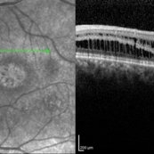
OCT image of the left eye of a 30-year-old man with juvenile retinoschisis. VA OS is 20/100.
Photographer: Shabnam Pooreh, Negah Eye Center, Tehran , Iran
Imaging device: Specteralis
Condition/keywords: juvenile retinoschisis, macular schisis, optical coherence tomography (OCT)
-
 Juvenile Retinoschisis
Oct 10 2015 by Hamid Ahmadieh, MD
Color fundus photograph of the right eye of a 30-year-old man with juvenile retinoschisis and typical macular changes.
Photographer: Shabnam Pooreh, Negah Eye Center, Tehran , Iran
Condition/keywords: color fundus photograph, juvenile retinoschisis, macular schisis
-
 Juvenile Retinoschisis
Oct 10 2015 by Hamid Ahmadieh, MD
OCT image of the left eye of a 30-year-old man with juvenile retinoschisis. VA OS is 20/100.
Photographer: Shabnam Pooreh, Negah Eye Center, Tehran , Iran
Imaging device: Specteralis
Condition/keywords: juvenile retinoschisis, macular schisis, optical coherence tomography (OCT)
-
 Optic Pit 1
Oct 10 2013 by Roy Schwartz, MD
Young patient with optic pit and macular schisis.
Photographer: Galit Yair Pur
-
 Juvenile Retinoschisis
Oct 10 2015 by Hamid Ahmadieh, MD
OCT image of the left eye of a 30-year-old man with juvenile retinoschisis. VA OS is 20/100.
Photographer: Shabnam Pooreh, Negah Eye Center, Tehran , Iran
Imaging device: Specteralis
Condition/keywords: juvenile retinoschisis, macular schisis, optical coherence tomography (OCT)
-
 Juvenile Retinoschisis
Oct 10 2015 by Hamid Ahmadieh, MD
OCT image of the left eye of a 30-year-old man with juvenile retinoschisis. VA OS is 20/100.
Photographer: shabnam Pooreh, Negah Eye Center, Tehran , Iran
Imaging device: Specteralis
Condition/keywords: juvenile retinoschisis, macular schisis, optical coherence tomography (OCT)
-
 Lamellar Macular Hole From Myopic Schisis
Aug 26 2018 by John T. Thompson, MD
Spectral domain OCT of myopic patient presenting with a lamellar macular hole and surrounding macular schisis.
Imaging device: Heidelberg Spectralis
Condition/keywords: lamellar macular hole, myopic macular schisis
-
 Myopic Traction Maculopathy
Mar 17 2025 by Drew Mitchell
HD 1 line 100x 9 mm scan of a right eye with MTM at stage 3c. Macular Schisis Detachment.
Photographer: Drew Mitchell OCT-C
Imaging device: Zeiss Cirrus 5000
Condition/keywords: full thickness macular hole, Macular hole, myopic foveoschisis, myopic macular schisis, myopic traction maculopathy, PVD
-
 Macular Schisis with RD
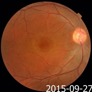
Apr 4 2025 by Tejaswita Verma
Fundus photo of a 29 year-old male with RRD, breaks in periphery, macular schisis . Vision CF2.5 mts
Photographer: DR. TEJASWITA VERMA
Imaging device: MIRANTE
Condition/keywords: macular schisis, retinal detachment
 Loading…