Search results (64 results)
-
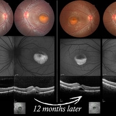 Vitelliform Macular Dystrophy or Best Disease
Dec 16 2016 by Young Hee Yoon, MD, PhD
Bilateral fundus photographs and autofluorescence images of 15-year-old girl who was diagnosed as vitelliform macular dystrophy or Best disease. Vitelliform macular lesion showed morphologic change during one year.
Photographer: Hyejin Jo, Sunghyun Kim, Heoni Hong, Minjung Chae, Mihwa Shin, Asan medical center, Seoul
Imaging device: Topcon TRC-500X fundus camera, Heidelberg HRA 2 autofluorescence, Heldelberg Spectralis OCT
Condition/keywords: Best disease, pseudohypopyon, scrambled-egg, vitelliform macular dystrophy
-
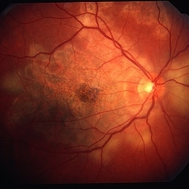
 Retinoblastoma - Regressed
May 3 2013 by Suber S. Huang, MD, MBA, FASRS
24-year-old male status post radiation for retinoblastoma with secondary metastatic carcinoma.
Imaging device: Retina Diseases Imaging Analysis Reading Center
Condition/keywords: endophytic tumor growth, intraocular tumor, macular lesion, radiotherapy, retinoblastoma
-
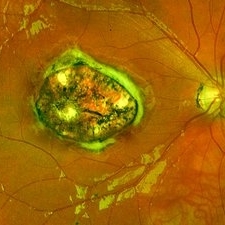
 Serpiginous Choroiditis
Feb 25 2013 by Henry J. Kaplan, MD
Serpiginous choroiditis, right eye. Both active and inactive lesions clearly visible; active lesions are the yellowish subretinal area most prominant nasal to optic nerve head and also around the inferior arcade and temporal to the macular lesion.
Condition/keywords: serpiginous choroiditis
-
---thumb.jpg/image-square;max$300,300.ImageHandler) Congenital Toxoplasmosis
Congenital Toxoplasmosis
Aug 13 2013 by From the Collections of Thomas M. Aaberg, MD and Thomas M. Aaberg Jr., MD
Hyper and hypo oimentedmacular lesion.
Condition/keywords: congenital toxoplasmosis
-
 Presumed Ocular Histoplasmosis
Jul 11 2013 by Jerald A. Bovino, MD
No history, macular lesion with hemorrhage.
Condition/keywords: ocular histoplasmosis syndrome (OHS)
-
---thumb.jpg/image-square;max$300,300.ImageHandler) Congenital Toxoplasmosis
Congenital Toxoplasmosis
Aug 13 2013 by From the Collections of Thomas M. Aaberg, MD and Thomas M. Aaberg Jr., MD
Hyper and hypo oimentedmacular lesion.
Condition/keywords: congenital toxoplasmosis
-
---thumb.jpg/image-square;max$300,300.ImageHandler) Right Macular Lesion
Right Macular Lesion
Feb 3 2014 by Maurice F. Rabb
Male patient with lesion in the right macula. It appears he has a small area of epiretinal or intraretinal fibrotic or fibrotic tissue wiht nonperfusion encircled by telangienctasis.
Condition/keywords: macular lesion, retinal telangiectasia
-
 Central Macular Lesion, Choroidal Hemangioma, Staphyloma
Jul 11 2013 by Jerald A. Bovino, MD
No history.
Condition/keywords: central vascular lesion, staphyloma
-
 X-linked Juvenile Retinoschisis
Feb 20 2015 by H. Michael Lambert, MD
No history. Hyperpigmented central macular lesion in the right eye. Difficult to tell if there is inferior peripheral schisis into the posterior pole. Some sheathing of vessels coursing inferiorly from the optic nerve
Condition/keywords: juvenile retinoschisis
-
---thumb.jpg/image-square;max$300,300.ImageHandler) Congenital Toxoplasmosis
Congenital Toxoplasmosis
Aug 13 2013 by From the Collections of Thomas M. Aaberg, MD and Thomas M. Aaberg Jr., MD
Hyper and hypo oimentedmacular lesion.
Condition/keywords: congenital toxoplasmosis
-
---thumb.jpg/image-square;max$300,300.ImageHandler) Congenital Toxoplasmosis
Congenital Toxoplasmosis
Aug 13 2013 by From the Collections of Thomas M. Aaberg, MD and Thomas M. Aaberg Jr., MD
Hyper and hypo oimentedmacular lesion.
Condition/keywords: congenital toxoplasmosis
-
---thumb.jpg/image-square;max$300,300.ImageHandler) Congenital Toxoplasmosis
Congenital Toxoplasmosis
Aug 13 2013 by From the Collections of Thomas M. Aaberg, MD and Thomas M. Aaberg Jr., MD
Hyper and hypo oimentedmacular lesion.
Condition/keywords: congenital toxoplasmosis
-
---thumb.jpg/image-square;max$300,300.ImageHandler) Congenital Toxoplasmosis
Congenital Toxoplasmosis
Aug 13 2013 by From the Collections of Thomas M. Aaberg, MD and Thomas M. Aaberg Jr., MD
Hyper and hypo oimentedmacular lesion.
Condition/keywords: congenital toxoplasmosis
-
 Congenital Toxoplasmosis
Dec 18 2019 by Yoshihiro Yonekawa, MD, FASRS
Widefield fundus image of a teenage girl's right eye with an inactive congenital toxoplasmosis macular lesion. Her vision is 20/400 in this eye.
Photographer: Netanya Lerner, COA, Wills Eye Hospital/Mid Atlantic Retina
Imaging device: Optos California
Condition/keywords: congenital toxoplasmosis, pediatric retina
-
---thumb.jpg/image-square;max$300,300.ImageHandler) Congenital Toxoplasmosis
Congenital Toxoplasmosis
Aug 13 2013 by From the Collections of Thomas M. Aaberg, MD and Thomas M. Aaberg Jr., MD
Hyper and hypo oimentedmacular lesion.
Condition/keywords: congenital toxoplasmosis
-
---thumb.jpg/image-square;max$300,300.ImageHandler) Congenital Toxoplasmosis
Congenital Toxoplasmosis
Aug 13 2013 by From the Collections of Thomas M. Aaberg, MD and Thomas M. Aaberg Jr., MD
Hyper and hypo oimentedmacular lesion.
Condition/keywords: congenital toxoplasmosis
-
---thumb.jpg/image-square;max$300,300.ImageHandler) Congenital Toxoplasmosis
Congenital Toxoplasmosis
Aug 13 2013 by From the Collections of Thomas M. Aaberg, MD and Thomas M. Aaberg Jr., MD
Hyper and hypo oimentedmacular lesion.
Condition/keywords: congenital toxoplasmosis
-
---thumb.JPG/image-square;max$300,300.ImageHandler) Exudative AMD
Exudative AMD
Dec 9 2012 by Mallika Goyal, MD
Left eye fundus shows resolving macular exudates following a year of anti-VEGF therapy and PDT in a patient with bilateral advanced AMD with large macular lesions and gravitational exudates.
Photographer: Mallika Goyal, MD, Apollo Health City, Jubilee Hills, Hyderabad
Condition/keywords: exudative age-related macular degeneration
-
---thumb.jpg/image-square;max$300,300.ImageHandler) Congenital Toxoplasmosis
Congenital Toxoplasmosis
Aug 13 2013 by From the Collections of Thomas M. Aaberg, MD and Thomas M. Aaberg Jr., MD
Hyper and hypo oimentedmacular lesion.
Condition/keywords: congenital toxoplasmosis
-
 Peripapillary Scarring Associated With Atrophic Macular Lesion (Resolution)
Jul 11 2013 by Jerald A. Bovino, MD
No history, lesion resolved, perhaps post surgery.
Condition/keywords: hemorrhagic macular disciform lesion resolved
-
---thumb.jpg/image-square;max$300,300.ImageHandler) Congenital Toxoplasmosis
Congenital Toxoplasmosis
Aug 13 2013 by From the Collections of Thomas M. Aaberg, MD and Thomas M. Aaberg Jr., MD
Hyper and hypo oimentedmacular lesion.
Condition/keywords: congenital toxoplasmosis
-
 X-linked Juvenile Retinoschisis
Feb 20 2015 by H. Michael Lambert, MD
No history. Hyperpigmented central macular lesion in the right eye. Difficult to tell if there is inferior peripheral schisis into the posterior pole.
Condition/keywords: juvenile retinoschisis
-
---thumb.jpg/image-square;max$300,300.ImageHandler) Congenital Toxoplasmosis
Congenital Toxoplasmosis
Aug 13 2013 by From the Collections of Thomas M. Aaberg, MD and Thomas M. Aaberg Jr., MD
Hyper and hypo oimentedmacular lesion.
Condition/keywords: congenital toxoplasmosis
-
---thumb.jpg/image-square;max$300,300.ImageHandler) Congenital Toxoplasmosis
Congenital Toxoplasmosis
Aug 13 2013 by From the Collections of Thomas M. Aaberg, MD and Thomas M. Aaberg Jr., MD
Hyper and hypo oimentedmacular lesion.
Condition/keywords: congenital toxoplasmosis
 Loading…