Search results (62 results)
-
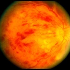
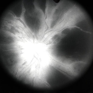
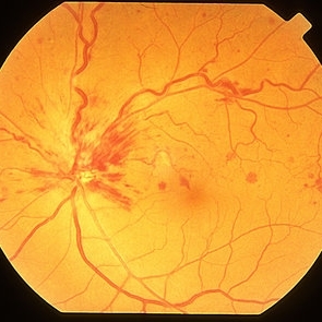 Non Ischemic Hemi-CRVO
Mar 29 2013 by Henry J. Kaplan, MD
Non-ischemic CRVO: blurred disc margins, dilated and tortous veins and scattered hemorrhages in the superior half of the retina.
Condition/keywords: branch retinal vein occlusion (BRVO), central retinal vein occlusion (CRVO), hemi CRVO, non-ischemic central retinal vein occlusion (CRVO)
-
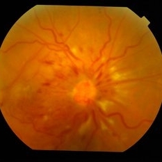
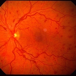 Non Ischemic CRVO
Mar 29 2013 by Henry J. Kaplan, MD
Non ischemic CRVO.
Condition/keywords: central retinal vein occlusion (CRVO)
-
 Angle Neovascularization
Mar 21 2013 by Yusuke Oshima, MD, PhD
Angle neovascularization due to ischemic CRVO.
Photographer: Yusuke Oshima, MD, PhD, Osaka University Graduate School of Medicine
Condition/keywords: angle neovascularization, gonioscopy
-
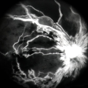
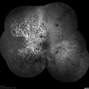
 CRVO ischemic - FA 1
Jan 11 2013 by Alex P. Hunyor, MD
Severely ischaemic central retinal vein obstruction (CRVO), right eye - early fluorescein angiogram showing almost complete capillary non-perfusion.
Condition/keywords: ischemic CRVO
-
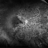
 CRVO ischemic - FA 2
Jan 11 2013 by Alex P. Hunyor, MD
Severely ischaemic central retinal vein obstruction (CRVO), right eye - mid phase fluorescein angiogram.
Condition/keywords: ischemic CRVO
-
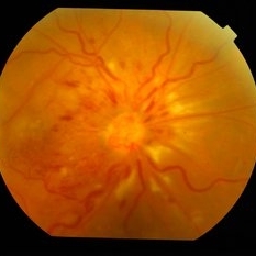
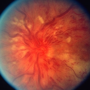 CRVO
CRVO
Mar 29 2013 by Henry J. Kaplan, MD
Full blown ischemic CRVO with disc swelling, dilated and tortous veins, scattered hemorrhages and multiple cotton wool spots.
Condition/keywords: central retinal vein occlusion (CRVO), ischemic CRVO
-
---thumb.JPG/image-square;max$300,300.ImageHandler) Ischaemic CRVO
Ischaemic CRVO
Jan 28 2014 by Mallika Goyal, MD
Left fundus of a 65-year-old diabetic and hypertensive gentleman shows ischaemic CRVO with extensive retinal and pre-retinal hemorrhage, severe macular edema but no neovascularisation as confirmed by fluorescein angiography.
Photographer: Mallika Goyal, MD, Apollo Health City, Hyderabad, India
Condition/keywords: ischemic CRVO
-
 CRVO ischemic - color image
Jan 11 2013 by Alex P. Hunyor, MD
Severely ischaemic central retinal vein obstruction (CRVO), right eye - color image.
Condition/keywords: central retinal vein occlusion (CRVO), ischemic CRVO
-
 CRVO With NVD
May 15 2014 by Mallika Goyal, MD
Right fundus of a 50-year-old gentleman shows NVD and inferior settled vitreous heme signifying ischemic conversion of an initially non-ischemic CRVO.
Photographer: Mallika Goyal, MD, Apollo Health City, Jubilee Hills, Hyderabad, India
Condition/keywords: ischemic CRVO
-
---thumb.JPG/image-square;max$300,300.ImageHandler) Ischaemic CRVO
Ischaemic CRVO
Jan 28 2014 by Mallika Goyal, MD
Left fundus of a 65-year-old diabetic and hypertensive gentleman shows ischaemic CRVO with extensive retinal and pre-retinal haemorrhage, severe macular edema but no neovascularisation as confirmed by fluorescein angiography.
Photographer: Mallika Goyal, MD, Apollo Health City, Hyderabad, India
Condition/keywords: ischemic CRVO
-
---thumb.JPG/image-square;max$300,300.ImageHandler) Ischaemic CRVO
Ischaemic CRVO
Jan 28 2014 by Mallika Goyal, MD
Left fundus fluorescein angiogram of a 65-year-old diabetic and hypertensive gentleman shows ischaemic CRVO with no neovascularization.
Photographer: Mallika Goyal, MD, Apollo Health City, Hyderabad, India
Condition/keywords: ischemic CRVO
-
 CRVO ischemic - FA 3
Jan 11 2013 by Alex P. Hunyor, MD
Severely ischaemic central retinal vein obstruction (CRVO), right eye - late phase fluorescein angiogram.
Condition/keywords: ischemic CRVO
-
---thumb.JPG/image-square;max$300,300.ImageHandler) Ischaemic CRVO
Ischaemic CRVO
Jan 28 2014 by Mallika Goyal, MD
Left fundus of a 65-year-old diabetic and hypertensive gentleman shows ischaemic CRVO with extensive retinal and pre-retinal hemorrhage, severe macular edema but no neovascularisation as confirmed by fluorescein angiography.
Photographer: Mallika Goyal, MD, Apollo Health City, Hyderabad, India
Condition/keywords: ischemic CRVO
-
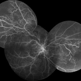
 Central Retinal Vein Occlusion with Severe Ischemia
May 22 2016 by Olivia Rainey
Composite fluorescein angiogram of the left eye of a male with a Central Retinal Vein Occlusion with severe ischemia.
Photographer: Olivia Rainey
Imaging device: Heidelberg Spectralis
Condition/keywords: central retinal vein occlusion (CRVO), composite, fluorescein leakage, ischemic CRVO
-
 Ischemic Central Retinal Vein Occlusion
Jan 24 2019 by Nichole Lewis
76-year-old woman with an ischemic central retinal vein occlusion, severely attenuated and sclerotic vessels and scattered retinal hemorrhages. Vision decrease over 1 year. VA 20/CF. Patient is returning for pan retinal photocoagulation.
Photographer: Nichole Lewis
Imaging device: Optos
Condition/keywords: attenuated vessels, central retinal vein occlusion (CRVO), hemorrhage, ischemic CRVO, sclerotic vessels
-
 CRVO With NVD
May 15 2014 by Mallika Goyal, MD
Right fundus of a 50-year-old gentleman shows NVD and inferior settled vitreous heme signifying ischemic conversion of an initially non-ischemic CRVO.
Photographer: Mallika Goyal, MD, Apollo Health City, Jubilee Hills, Hyderabad, India
Condition/keywords: ischemic CRVO
-
---thumb.JPG/image-square;max$300,300.ImageHandler) Ischaemic CRVO
Ischaemic CRVO
Jan 28 2014 by Mallika Goyal, MD
Left fundus fluorescein angiogram of a 65-year-old diabetic and hypertensive gentleman shows ischaemic CRVO with no neovascularization.
Photographer: Mallika Goyal, MD, Apollo Health City, Hyderabad, India
Condition/keywords: ischemic CRVO
-
 CRVO With NVD
May 15 2014 by Mallika Goyal, MD
Right fundus of a 50-year-old gentleman shows NVD and inferior settled vitreous heme signifying ischemic conversion of an initially non-ischaemic CRVO.
Photographer: Mallika Goyal, MD, Apollo Health City, Jubilee Hills, Hyderabad, India
Condition/keywords: ischemic CRVO
-
 CRVO with Secondary CLRAO
May 28 2020 by Richard M Martindale, MD
Non-ischemic CRVO (VA 20/30) with secondary CLRAO (nasal macular pallor) in a hypertensive 69yo female. Pathophysiologically, the cilioretinal artery occlusion occurs secondary to elevation in the hydrostatic pressure in the retinal venous system relative to the choroidal perfusion pressure (which supplies the cilioretinal artery).
Photographer: Retina Consultants of Alabama, P. C.
Imaging device: Optos
Condition/keywords: cilioretinal artery occlusion, non-ischemic central retinal vein occlusion (CRVO)
-
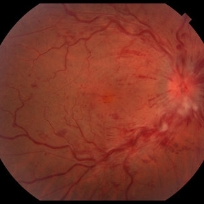
 Ischemic CRVO
Jul 28 2018 by Carlos Quezada-Ruiz, MD, FASRS
Fundus photograph of a 75-year-old woman with hypertension and dyslipidemia who presented to the clinic with sudden decrease in vision on her right eye.
Condition/keywords: central retinal vein occlusion (CRVO)
-
 CRVO
CRVO
-
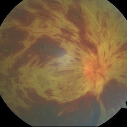
 Central Retinal Vein Occlusion (CRVO) Associated with Papillophlebitis
Apr 16 2021 by Gabriel Costa Andrade, PhD
Fundus photograph of an 38-year-old woman with Central retinal vein occlusion (CRVO) associated with papillophlebitis.
Photographer: Dr Gabriel Andrade
Condition/keywords: central retinal vein occlusion (CRVO), ischemic CRVO
-
 Central Retinal Vein Occlusion
Dec 22 2018 by FELIPE PEREIRA
25-year-old male patient with acute and painless vision loss of left eye. The fundus examination demonstrate optic disc swelling, venous tortuosity, diffuse intraretinal hemorrhage and severe macular edema. There is also extensive exudative retinal detachment with lipid deposits in the posterior pole, mainly around the vessels. The systemic work up was negative, including serologies, rheumatologic and hematological markers and cholesterol and triglycerides within normal limits.
Photographer: Felipe Pereira
Imaging device: Vizucan, Zeiss
Condition/keywords: central vein occlusion, ischemic CRVO
 Loading…