Search results (14 results)
-
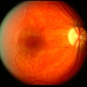
 Siegrist Streaks
Mar 29 2013 by Henry J. Kaplan, MD
Typical Siegrist streaks in hypertensive choridopathy; hyperpigmentations in a linear fashion along choroidal vessels , a rare finding.
Condition/keywords: hypertensive choroidopathy, Siegrist Streaks
-
 Hypertensive Choroidopathy - Right Eye
Dec 21 2016 by Maciej Czepita
Fundus photograph and SD-OCT scan as well as fundus autofluorescence image (FAF) of the right eye of a 70-year-old woman with hypertensive choroidopathy. In the fundus image numerous Elschnig's spots are visible. Note the Hollenhorst plaque in the superior temporal artery. In the SD-OCT scan (green line on the fundus image) the RPE layer is uneven. Numerous hypo and hyperautofluorescent patches can be seen in the fundus autofluorescence image.
Photographer: Maciej Czepita, M.D., Ph.D., Pomeranian Medical University, Szczecin, Poland
Imaging device: Heidelberg Spectralis HRA+OCT
Condition/keywords: hypertensive choroidopathy
-
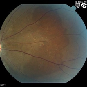
 Hypertensive Choroidopathy
Dec 7 2012 by F. Ryan Prall, MD
32-year-old male with decreased vision, admitted for malignant hypertension.
Photographer: Tom Egnatz, Indiana University
Condition/keywords: hypertensive choroidopathy, malignant hypertension
-
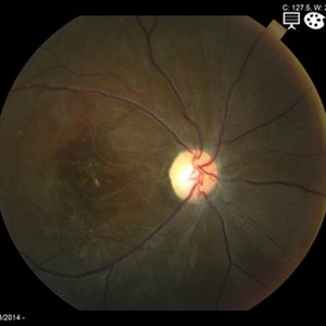
 Hypertensive optic neuropathy and choroidopathy right eye
Jan 11 2013 by Alex P. Hunyor, MD
Previous hypertensive optic neuropathy and choroidopathy, right eye. A young female who had a history severe pre-eclampsia. Note optic atrophy and multiple Elschnig spots.
Condition/keywords: hypertensive choroidopathy, hypertensive optic neuropathy
-
 Hypertensive optic neuropathy and choroidopathy left eye
Jan 11 2013 by Alex P. Hunyor, MD
Previous hypertensive optic neuropathy and choroidopathy, right eye. A young female who had a history severe pre-eclampsia. Note optic atrophy and multiple Elschnig spots.
Condition/keywords: hypertensive choroidopathy, hypertensive optic neuropathy
-
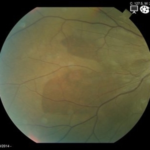
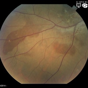 Choroidal Hemorrhage from Hypertensive Choroidopathy
May 19 2014 by Charline Boente
Fundus photograph of a 21-year-old African-American female with HTN and end-stage renal disease from IgA nephropathy.
Photographer: Mark Harrod, University Hospitals Eye Institute, Case Western Reserve University
Condition/keywords: hypertensive choroidopathy
-
 Hypertensive Choroidopathy - Left Eye
Dec 21 2016 by Maciej Czepita
Fundus photograph, SD-OCT image and fundus autofluorescence image (FAF) of a 70-year-old woman with hypertensive choroidopathy. Multiple Elchnig's spots can be seen as well as two Siegrist's spots near the superior temporal artery and inferotemporal vein. The RPE layer is uneven in the SD-OCT scan (green line on the fundus image shows the location of the scan). Numerous hypo and hyperautofluorescent patches are seen in the fundus autofluorescence image.
Photographer: Maciej Czepita, M.D., Ph.D., Pomeranian Medical University, Szczecin, Poland
Imaging device: Heidelberg Spectralis HRA+OCT
Condition/keywords: hypertensive choroidopathy
-
 Choroidal Hemorrhage from Hypertensive Choroidopathy
May 19 2014 by Charline Boente
Fundus photograph of a 21-year-old African-American female with HTN and end-stage renal disease from IgA nephropathy.
Photographer: Mark Harrod, University Hospitals Eye Institute, Case Western Reserve University
Condition/keywords: hypertensive choroidopathy
-
 Hypertensive Retinopathy, Right
Feb 23 2017 by Alla Goldberg, MD
Fundus photograph of 35-year-old man with severe hypertension (182/128).
Photographer: Sofia Rutiaga, UT Health McGovern Medical School, Cizik Eye Clinic
Condition/keywords: cotton wool spots, Elschnig's spots, hypertensive choroidopathy, hypertensive retinopathy, serous retinal detachment
-
 Choroidal Hemorrhage from Hypertensive Choroidopathy
May 19 2014 by Charline Boente
Fundus photograph of a 21-year-old African-American female with HTN and end-stage renal disease from IgA nephropathy.
Photographer: Mark Harrod, University Hospitals Eye Institute, Case Western Reserve University
Condition/keywords: hypertensive choroidopathy
-
 Hypertensive retinopathy, left
Feb 23 2017 by Alla Goldberg, MD
Fundus photograph of 35-year-old man with severe hypertension (182/128).
Photographer: Sofia Rutiaga, UT Health McGovern Medical School, Cizik Eye Clinic
Condition/keywords: cotton wool spots, Elschnig's spots, hypertensive choroidopathy, hypertensive retinopathy, serous retinal detachment
-
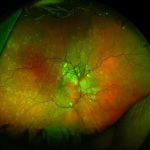
 Blistered Retina
Jan 27 2024 by prathibha hande, MS DNB
Fundus photo of a 32 year old male presenting with blurred vision. Undiagnosed renal hypertension. Blood pressure at the time of presentation 210/120 mmhg.
Photographer: Mr Prathap K
Imaging device: Mirante SLO fundus camera
Condition/keywords: hypertensive choroidopathy
-
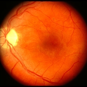
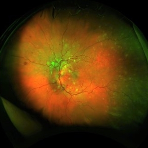
 Siegrist Streaks and Hypertensive Choroidopathy
Jul 29 2023 by Júlio Costa Almeida, MD
Fundus photograph of a 68-year-old woman with hypertensive retinopathy, choroidopathy and the classic Siegrist streaks.
Imaging device: Nonmyd 8s by Kowa
Condition/keywords: hypertensive choroidopathy, hypertensive retinopathy, Siegrist Streaks
 Loading…